Coordination and Movement
3. Muscles and Movement
Learning outcomes
- I can explain how muscles produce movement.
- I can describe the interaction between muscles and bones.
- I can distinguish between different types of muscle tissue.
- I can explain the role of energy in muscle contraction.
- I can analyze how movement supports survival and behavior.
How Do Animals Produce Movement?
Movement is one of the most obvious characteristics of animals. Animals move their entire bodies to find food, escape predators, locate mates, and explore their environments. They also produce smaller movements involved in breathing, digestion, circulation, and communication.
In vertebrates, most visible body movement results from cooperation between three major systems:
- The nervous system sends signals that control muscles.
- The muscular system produces force by contracting.
- The skeletal system provides support and structures that muscles can pull against.
Together, the muscles and skeleton form the musculoskeletal system.


A useful sequence is:
Nervous signal → muscle contraction → force on bone → movement at joint
Muscles Produce Force by Contracting
Muscles produce movement through contraction.
When a muscle contracts, it develops tension and can shorten, pulling on structures attached to it.
An important principle is:
Muscles pull; they do not actively push.
This means that moving a joint in opposite directions usually requires different muscles.
For example, bending and straightening the elbow involve different muscles working together.
Muscles and Bones
Most skeletal muscles are attached to bones by strong connective tissues called tendons.
When a skeletal muscle contracts:
- The muscle produces tension.
- The tendon transmits the force.
- The force pulls on a bone.
- The bone moves around a joint.


The skeleton therefore does more than support the body. It provides a mechanical framework against which muscles can produce movement.
Tendons and Ligaments
Tendons and ligaments are often confused.
A tendon connects:
Muscle → bone
A ligament connects:
Bone → bone
Ligaments help stabilise joints, while tendons transfer muscular force to the skeleton.
Joints
A joint is a location where two or more bones meet.
Some joints permit little or no movement, while others allow considerable movement.
Examples of movable joints include:
- Shoulder.
- Elbow.
- Wrist.
- Hip.
- Knee.
- Ankle.


Many freely movable joints are called synovial joints.
They contain structures that reduce friction and allow controlled movement.
Bones Act as Levers
Bones can act as levers.
A lever is a rigid structure that rotates around a pivot.
In the body:
- Bone acts as the lever.
- Joint acts as the pivot.
- Muscle provides the force.
- Body part or external object provides resistance.
For example, when the biceps contracts to lift the forearm, the forearm bones rotate around the elbow joint.
This arrangement converts muscle contraction into useful movement.
Antagonistic Muscle Pairs
Because muscles pull rather than push, many skeletal muscles work in antagonistic pairs.
An antagonistic pair consists of muscles that produce opposite movements.
The biceps and triceps are a familiar example.


Bending the Arm
When the elbow bends:
- Biceps contracts.
- Biceps shortens and produces tension.
- Triceps relaxes sufficiently to allow movement.
- Forearm moves upward.
This movement is called flexion.
The biceps acts as a flexor at the elbow.
Straightening the Arm
When the elbow straightens:
- Triceps contracts.
- Triceps produces force on the forearm.
- Biceps relaxes sufficiently to allow movement.
- Forearm moves downward or away from the upper arm.
This movement is called extension.
The triceps acts as an extensor at the elbow.
Therefore:
Flexion → biceps contracts
Extension → triceps contracts
In real movements, muscle control can be more complex, and opposing muscles may sometimes contract simultaneously to stabilise a joint.
Muscle Tissue
Animals contain different types of muscle tissue specialised for different functions.
Humans have three major types:
- Skeletal muscle.
- Smooth muscle.
- Cardiac muscle.


Although all three types can contract, their structures, locations, and functions differ.
Skeletal Muscle
Skeletal muscle is usually attached to bones.
It produces movements such as:
- Walking.
- Running.
- Jumping.
- Writing.
- Chewing.
- Moving the eyes.
- Maintaining posture.
Skeletal muscle is usually described as voluntary muscle because many of its movements can be consciously controlled.
However, skeletal muscles are also involved in automatic responses such as reflexes.
Under a microscope, skeletal muscle has a striped or striated appearance.
Smooth Muscle
Smooth muscle is found in the walls of many internal organs.
Examples include:
- Digestive tract.
- Blood vessels.
- Bladder.
- Airways.
- Uterus.
Smooth muscle usually operates without conscious control and is therefore described as involuntary.


For example, smooth muscle in the digestive tract produces waves of contraction called peristalsis, which help move food through the digestive system.
Cardiac Muscle
Cardiac muscle is found only in the heart.
It contracts rhythmically throughout life to pump blood around the body.
Cardiac muscle is:
- Involuntary.
- Striated.
- Highly resistant to fatigue under normal conditions.
- Specialised for repeated rhythmic contraction.


Cardiac muscle cells are connected in ways that allow coordinated contraction of heart tissue.
Comparing Muscle Types
| Feature | Skeletal Muscle | Smooth Muscle | Cardiac Muscle |
|---|---|---|---|
| Main location | Attached to skeleton | Internal organs | Heart |
| Control | Mostly voluntary | Involuntary | Involuntary |
| Appearance | Striated | Non-striated | Striated |
| Typical role | Body movement | Movement within organs | Pumping blood |
| Contraction | Can be rapid and powerful | Usually slower | Rhythmic |
Each muscle type is adapted to its particular function.
Structure of Skeletal Muscle
A skeletal muscle is organised into increasingly smaller structures.
A simplified organisation is:
Whole muscle → muscle bundles → muscle fibres → myofibrils → contractile proteins
A muscle fibre is actually a specialised muscle cell.


Inside the fibres are many myofibrils, which contain the protein structures responsible for contraction.
Actin and Myosin
Two important proteins involved in muscle contraction are:
- Actin
- Myosin
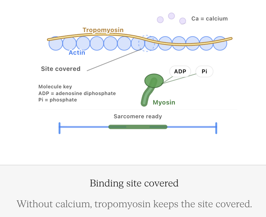
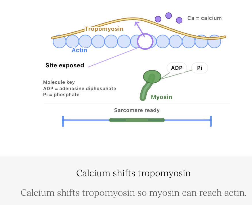
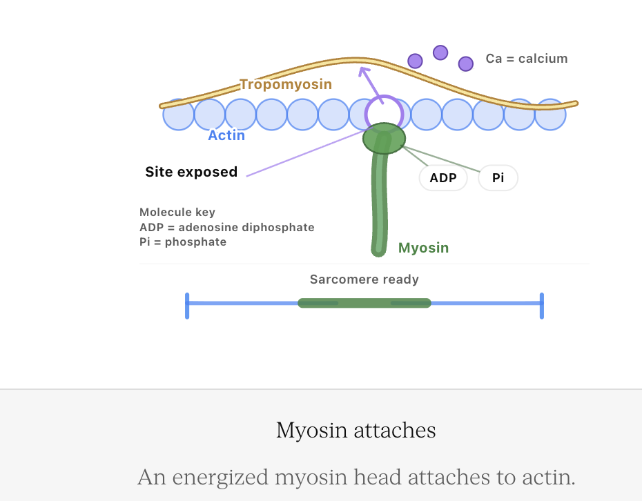
These proteins are arranged in repeating units called sarcomeres.
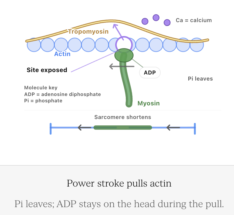
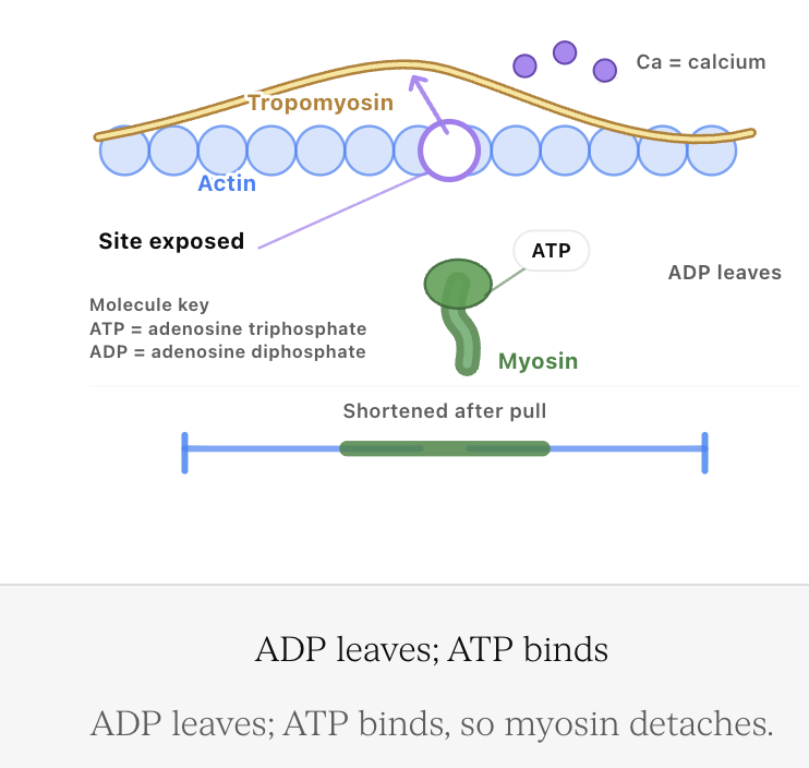
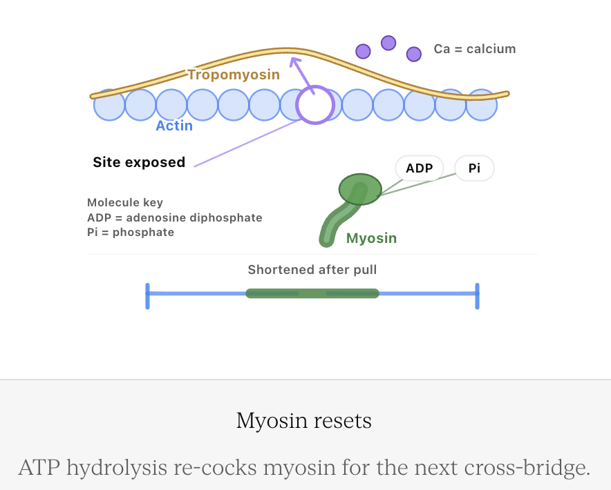
During contraction, actin and myosin interact so that the protein filaments slide past one another.
This is known as the sliding filament mechanism.


The filaments themselves do not simply become shorter.
Instead, their arrangement changes as they slide relative to one another, shortening the sarcomere.
Many sarcomeres shortening together causes the muscle fibre to shorten.
How Muscle Contraction Occurs
A skeletal muscle does not contract spontaneously during normal movement. It receives instructions from the nervous system.
A simplified sequence is:
Motor neuron stimulated
↓
Signal reaches muscle fibre
↓
Calcium ions become available inside the muscle fibre
↓
Actin and myosin interact
↓
Myosin pulls actin
↓
Sarcomeres shorten
↓
Muscle develops tension and may shorten