Coordination and Movement
| サイト: | Young Education |
| コース: | Animal Physiology |
| ブック: | Coordination and Movement |
| 印刷者: | 访客用户 |
| 日付: | 2026年 10月 5日(月曜日) 04:59 |
1. Nervous Systems
Learning outcomes
- I can identify the major components of a nervous system.
- I can explain how nerve cells transmit information.
- I can describe the functions of the brain, spinal cord, and nerves.
- I can explain how nervous systems coordinate responses.
- I can compare simple and complex nervous systems.
What Is a Nervous System?
Animals must constantly detect changes in their surroundings and inside their bodies.
They may need to:
- Detect light, sound, temperature, pressure, or chemicals.
- Locate food.
- Avoid predators.
- Maintain balance.
- Coordinate movement.
- Control internal organs.
- Respond rapidly to danger.
- Learn from previous experiences.
The nervous system is a communication and coordination system that detects information, processes it, and produces appropriate responses.
A simplified pathway is:
Stimulus → receptor → nervous system → effector → response


The nervous system is particularly important for rapid communication within an animal.
Major Components of the Nervous System
In humans and other vertebrates, the nervous system can be divided into two major parts:
- Central nervous system (CNS)
- Peripheral nervous system (PNS)
The central nervous system consists of:
- Brain
- Spinal cord
The peripheral nervous system consists mainly of:
- Nerves extending throughout the body.
Together, these structures allow information to travel between receptors, the central nervous system, and effectors.
The Central Nervous System
The central nervous system, or CNS, acts as a major processing and coordinating centre.
It consists of:
Brain + spinal cord
Information from sensory receptors can be sent to the CNS.
The CNS processes this information and can send signals to muscles or glands.


The Brain
The brain is an extremely complex organ containing billions of nerve cells and supporting cells.
Different regions perform different functions.
The brain is involved in:
- Processing sensory information.
- Coordinating movement.
- Memory.
- Learning.
- Emotions.
- Decision-making.
- Language.
- Maintaining balance.
- Controlling many automatic body functions.


The Cerebrum
The cerebrum is the largest region of the human brain.
It is involved in functions including:
- Conscious thought.
- Memory.
- Learning.
- Interpretation of sensory information.
- Voluntary movement.
- Language.
- Decision-making.
The outer region of the cerebrum, called the cerebral cortex, contains highly interconnected networks of neurons.
Different regions of the cortex are specialised for different functions, although they work together extensively.
The Cerebellum
The cerebellum is located toward the back and lower part of the brain.
It is especially important for:
- Coordination of movement.
- Balance.
- Posture.
- Fine control of muscular activity.
- Motor learning.
For example, catching a ball requires the nervous system to coordinate visual information with precise movements of the arms and hands.
The cerebellum contributes to this coordination.
The Brainstem
The brainstem connects the brain with the spinal cord.
It helps regulate several essential automatic functions, including aspects of:
- Breathing.
- Heart rate.
- Blood pressure.
- Swallowing.
- Sleep and wakefulness.
These functions continue without requiring conscious control.
This demonstrates that the nervous system coordinates both voluntary and involuntary activities.
The Spinal Cord
The spinal cord is a long bundle of nervous tissue extending from the brain through the vertebral column.
It performs two especially important functions.
Communication
It carries information between the brain and much of the body.
Coordination of Reflexes
It can coordinate some rapid responses without waiting for conscious processing by the brain.


The vertebral column surrounds and protects the spinal cord.
The Peripheral Nervous System
The peripheral nervous system, or PNS, connects the central nervous system with the rest of the body.
It includes nerves that carry information:
Toward the CNS
and
Away from the CNS
The PNS connects the brain and spinal cord with:
- Sensory receptors.
- Muscles.
- Glands.
- Internal organs.
Without the peripheral nervous system, the CNS would have no efficient way to receive information from or control most of the body.
What Is a Nerve?
A nerve is a bundle of nerve fibres, or axons, in the peripheral nervous system.
A useful comparison is an electrical cable.
A cable contains many individual wires.
Similarly, a nerve can contain many individual axons carrying signals.


A nerve and a neuron are therefore not the same thing.
A neuron is an individual cell.
A nerve contains many nerve fibres from neurons.
Neurons
The specialised cells that transmit information in the nervous system are called neurons.
A typical neuron contains:
- Cell body
- Dendrites
- Axon
- Axon terminals


The structure of a neuron is closely related to its function.
Dendrites
Dendrites are branching structures that receive signals from other cells.
Their branching shape provides a large surface area for connections.
A single neuron may receive information from many other neurons.
The Cell Body
The cell body contains:
- Nucleus.
- Cytoplasm.
- Organelles required to maintain the cell.
The cell body integrates many of the signals arriving at the neuron.
The Axon
The axon is a long extension that carries electrical signals away from the cell body.
Some axons are extremely long.
For example, neurons controlling muscles in the foot may have axons extending much of the length of the leg.
This allows rapid communication over relatively large distances.
Myelin
Many axons are surrounded by a fatty insulating material called the myelin sheath.
Myelin:
- Electrically insulates the axon.
- Helps signals travel more rapidly.
- Protects and supports the nerve fibre.


Small gaps between sections of myelin are called nodes of Ranvier.
In myelinated neurons, electrical activity effectively moves rapidly from node to node, greatly increasing conduction speed.
How Neurons Transmit Information
Neurons transmit information using both electrical and chemical signals.
Along an axon, information travels as electrical changes in the neuron's membrane.
These rapid signals are called action potentials or nerve impulses.
At connections between neurons, information is usually transmitted chemically.
Therefore:
Within a neuron → mainly electrical signalling
Between many neurons → chemical signalling across synapses
The Nerve Impulse
At rest, there is an electrical difference across a neuron's cell membrane.
This results from unequal distributions of ions inside and outside the cell.
When a neuron is sufficiently stimulated, ion channels in the membrane open and the electrical condition of the membrane changes rapidly.
This produces an action potential.
The action potential travels along the axon.


The signal does not simply consist of electricity flowing through the neuron like current through a copper wire.
It results from controlled movements of ions across the neuron's membrane.
Synapses
Neurons usually do not directly touch one another.
The small junction between one neuron and another cell is called a synapse.
At many synapses, a tiny gap called the synaptic cleft separates the cells.


Neurotransmitters
When an action potential reaches an axon terminal:
- Chemical messengers called neurotransmitters are released.
- Neurotransmitters move across the synaptic cleft.
- They bind to receptors on the next cell.
- This changes the activity of the receiving cell.
The sequence can be simplified as:
Electrical signal → chemical signal → electrical response
Synapses allow neurons to form enormous communication networks.
Types of Neurons
Three useful functional categories are:
- Sensory neurons
- Relay neurons
- Motor neurons
Sensory Neurons
Sensory neurons carry information from receptors toward the central nervous system.
Receptors can detect stimuli such as:
- Light.
- Sound.
- Pressure.
- Temperature.
- Chemicals.
- Tissue damage.
For example:
Heat receptor in skin → sensory neuron → CNS
Relay Neurons
Relay neurons, also called interneurons, are located mainly within the central nervous system.
They connect neurons and help process information.
A relay neuron may receive signals from several neurons and pass information to other neurons.
Complex nervous systems contain enormous networks of interconnected relay neurons.
Motor Neurons
Motor neurons carry signals from the central nervous system toward effectors.
Effectors include:
- Muscles.
- Glands.
For example:
CNS → motor neuron → arm muscle contracts


Receptors and Effectors
A nervous response usually begins with a receptor.
A receptor detects a stimulus.
The response is carried out by an effector.
An effector may be:
- A muscle that contracts.
- A gland that releases a substance.
A useful general pathway is:
Stimulus → receptor → sensory neuron → CNS → motor neuron → effector → response
Worked Example: Catching a Ball
Imagine someone throws a ball toward you.
Stimulus
Light reflected from the moving ball enters your eyes.
Receptors
Photoreceptors in the retina detect the light.
Sensory Information
Signals travel toward the brain.
Processing
The brain interprets:
- Direction.
- Speed.
- Position.
Motor Output
Signals travel through motor pathways to skeletal muscles.
Effectors
Muscles in the arms and hands contract.
Response
Your hands move into position and catch the ball.
This entire process requires rapid coordination between sensory and motor systems.
Reflex Actions
Some responses must occur extremely quickly.
A reflex is a rapid, automatic response to a stimulus.
For example, touching a dangerously hot surface can trigger rapid withdrawal of the hand.


The Reflex Arc
A simplified reflex pathway is:
Stimulus
↓
Receptor
↓
Sensory neuron
↓
Relay neuron in spinal cord
↓
Motor neuron
↓
Effector
↓
Response
The spinal cord can coordinate the withdrawal response before conscious awareness of the pain is fully processed by the brain.
This reduces reaction time.
Worked Example: Touching a Hot Object
Suppose your finger touches a hot pan.
Step 1
Temperature and pain receptors are stimulated.
Step 2
Sensory neurons carry impulses toward the spinal cord.
Step 3
Neurons in the spinal cord process the information.
Step 4
Motor neurons carry impulses toward arm muscles.
Step 5
Muscles contract.
Step 6
Your hand moves away.
Information also travels to the brain, allowing you to become consciously aware of the pain and what happened.
Voluntary and Involuntary Responses
Nervous systems coordinate both voluntary and involuntary responses.
Voluntary Responses
Usually involve conscious control.
Examples include:
- Writing.
- Walking toward a door.
- Throwing a ball.
- Playing a musical instrument.
Involuntary Responses
Occur without deliberate conscious control.
Examples include:
- Changes in heart rate.
- Changes in pupil size.
- Digestive movements.
- Many reflexes.
Both require coordinated nervous-system activity.
The Autonomic Nervous System
Many internal organs are regulated by the autonomic nervous system.
This system helps control functions such as:
- Heart rate.
- Digestion.
- Pupil diameter.
- Activity of certain glands.
- Diameter of some blood vessels.
These processes usually occur without conscious control.
This allows the nervous system to continuously adjust internal conditions while conscious attention is focused elsewhere.
Nervous Coordination and Homeostasis
The nervous system plays an important role in homeostasis.
For example, when body temperature changes, receptors detect the change.
Information is processed in the brain, particularly in the hypothalamus.
Responses can then be coordinated.
If the body becomes too hot, responses may include:
- Increased sweating.
- Changes in blood flow near the skin.
If the body becomes too cold, responses can include:
- Shivering.
- Changes in blood flow near the skin.
The nervous system therefore connects:
Detection → coordination → response
Simple Nervous Systems
Not all animals possess brains and nervous systems as complex as those of vertebrates.
Some relatively simple animals have a nerve net.
A nerve net consists of interconnected neurons distributed through the body rather than concentrated into a large central brain.


Cnidarians such as hydra and jellyfish have nerve nets.
These systems can coordinate:
- Movement.
- Feeding.
- Responses to touch.
- Contractions of the body.
A complex brain is not required for every type of coordinated behaviour.
Increasing Centralisation
Many animals show greater centralisation of nervous tissue.
Instead of neurons being distributed relatively evenly, large numbers become concentrated into:
- Nerve cords.
- Ganglia.
- Brains.
A ganglion is a cluster of neuron cell bodies.
This concentration of nervous tissue allows more complex processing of information.
Nervous Systems in Invertebrates
Many invertebrates have highly capable nervous systems.
For example, insects typically have:
- A brain.
- A ventral nerve cord.
- Several ganglia.
- Sophisticated sensory organs.


These systems allow behaviours such as:
- Flight.
- Navigation.
- Communication.
- Feeding.
- Courtship.
- Escape responses.
Complex behaviour does not require a vertebrate-style nervous system.
Vertebrate Nervous Systems
Vertebrates generally have a highly centralised nervous system consisting of:
- Brain.
- Spinal cord.
- Extensive peripheral nerves.
The brain contains specialised regions and enormous networks of interconnected neurons.
This allows sophisticated:
- Sensory processing.
- Motor control.
- Learning.
- Memory.
- Behaviour.


Comparing Simple and Complex Nervous Systems
| Simple nervous system | More complex centralised nervous system |
|---|---|
| May contain a nerve net | Contains specialised processing centres |
| Limited centralisation | Strong centralisation |
| Relatively simple processing | Extensive information processing |
| Coordinates basic responses | Can coordinate highly complex responses |
| Example: hydra | Example: vertebrates |
However, "simple" does not mean ineffective.
A nervous system evolves according to the requirements of an animal's lifestyle and environment.
Nervous System Complexity and Lifestyle
Nervous systems are closely related to how animals interact with their environments.
A mobile predator, for example, may need to:
- Detect moving prey.
- Determine distance.
- Coordinate rapid movement.
- Maintain balance.
- Remember locations.
- Select between different behaviours.
These tasks require substantial information processing.
A relatively sedentary animal may face different sensory and coordination demands.
Nervous systems therefore reflect both evolutionary history and lifestyle.
Reaction Time
Reaction time is the time between detecting a stimulus and producing a response.
Reaction time depends on several processes:
Stimulus detection → nerve transmission → processing → motor transmission → muscle response
For example, catching a falling ruler requires:
- Visual receptors to detect movement.
- Sensory information to reach the CNS.
- The CNS to process the information.
- Motor signals to reach hand muscles.
- Muscles to contract.
Reaction time can be investigated experimentally using a simple ruler-drop test.
Why Nervous Responses Are Fast
Nervous-system communication can occur rapidly because:
- Electrical signals travel quickly along axons.
- Myelin increases conduction speed in many neurons.
- Synapses allow organised communication between cells.
- Neural pathways can connect receptors directly with appropriate processing centres.
- Reflex pathways can produce responses without waiting for conscious decision-making.
This makes nervous control particularly suitable for responses that must occur within fractions of a second.
Nervous and Hormonal Communication
Animals also communicate internally using hormones.
The nervous and endocrine systems both coordinate body functions, but they operate differently.
| Nervous communication | Hormonal communication |
|---|---|
| Uses neurons | Uses hormones |
| Signals travel along specific pathways | Hormones travel mainly through blood |
| Usually rapid | Often slower |
| Responses may be short-lived | Responses can be longer-lasting |
| Can target very specific cells | Hormones can circulate widely but affect cells with appropriate receptors |
The two systems frequently work together.
For example, the brain can stimulate hormonal responses during stressful situations.
Worked Example: Predator Detection
Imagine a deer detects a predator.
Its nervous system must rapidly:
- Detect the predator using sensory receptors.
- Transmit information to the CNS.
- Process the threat.
- Coordinate muscle activity.
- Adjust heart rate and breathing.
- Produce escape behaviour.
The nervous system therefore integrates information from many sources and coordinates multiple organs at the same time.
This ability can directly affect survival.
Worked Example: Damage to the Spinal Cord
Suppose the spinal cord is severely damaged.
The brain itself may still function normally, but communication between the brain and parts of the body can be disrupted.
Depending on the location and severity of the damage, this can interfere with:
- Sensory information reaching the brain.
- Motor signals reaching muscles.
- Some autonomic functions.
This demonstrates the spinal cord's critical role as a communication pathway between the brain and body.
Common Mistakes
Saying the Brain Is the Entire Nervous System
The nervous system includes the brain, spinal cord, nerves, neurons, and associated structures.
Confusing a Neuron With a Nerve
A neuron is an individual cell.
A peripheral nerve contains many nerve fibres.
Saying Nerve Impulses Are Simply Electricity Flowing Through Wires
Nerve impulses result from controlled movements of ions across neuronal membranes.
Saying Neurons Always Touch
Most neurons communicate across small junctions called synapses.
Thinking All Nervous Communication Is Electrical
Signals travel electrically along neurons, but communication across many synapses involves chemical neurotransmitters.
Thinking Reflexes Do Not Involve the CNS
Many reflexes involve the spinal cord, which is part of the central nervous system.
Thinking Reflexes Never Reach the Brain
A spinal reflex can begin before conscious processing, but information can still travel to the brain.
Assuming Simple Animals Have No Nervous System
Many simple animals have nerve nets or other forms of nervous organisation.
Assuming More Complex Always Means "Better"
Different nervous systems are adapted to different lifestyles and environments.
Check Your Understanding
1. What is the main function of a nervous system?
2. Name the two major divisions of the vertebrate nervous system.
3. What structures make up the central nervous system?
4. Describe two functions of the brain.
5. Explain two functions of the spinal cord.
6. What is the difference between a neuron and a nerve?
7. Describe the functions of dendrites and an axon.
8. Explain how myelin affects nerve transmission.
9. What happens at a synapse?
10. Compare sensory, relay, and motor neurons.
11. Write the pathway from a stimulus to a response using the terms receptor, CNS, sensory neuron, motor neuron, and effector.
12. Explain why withdrawal reflexes can protect an animal from injury.
13. Compare a nerve net with a centralised nervous system.
14. Explain how an animal's lifestyle might influence the complexity of its nervous system.
15. A person can feel a sharp object touching their foot but cannot voluntarily move the foot. What does this suggest about which nervous pathways may still be functioning and which may be disrupted?
Key Terms
- Nervous system – communication and coordination system that detects information, processes it, and coordinates responses.
- Central nervous system (CNS) – brain and spinal cord.
- Peripheral nervous system (PNS) – nerves connecting the CNS with the rest of the body.
- Brain – major processing and coordinating organ of the nervous system.
- Spinal cord – nervous tissue carrying information between the brain and body and coordinating many reflexes.
- Neuron – specialised cell that transmits information.
- Nerve – bundle of nerve fibres in the peripheral nervous system.
- Dendrite – branching part of a neuron specialised for receiving signals.
- Axon – long neuronal extension that carries electrical signals away from the cell body.
- Myelin sheath – insulating layer around many axons that increases conduction speed.
- Action potential – rapid electrical change that travels along a neuron's membrane.
- Synapse – junction through which a neuron communicates with another cell.
- Neurotransmitter – chemical messenger released at many synapses.
- Sensory neuron – neuron carrying information from receptors toward the CNS.
- Relay neuron – neuron within the CNS that connects and processes information between other neurons.
- Motor neuron – neuron carrying signals toward effectors.
- Receptor – specialised cell or structure that detects a stimulus.
- Effector – muscle or gland that produces a response.
- Reflex – rapid, automatic response to a stimulus.
- Reflex arc – neural pathway involved in producing a reflex.
- Nerve net – distributed network of neurons found in some relatively simple animals.
- Ganglion – cluster of neuron cell bodies.
- Centralisation – evolutionary concentration of nervous tissue into specialised processing regions.
- Reaction time – time between detecting a stimulus and producing a response.
Key Takeaways
- The nervous system allows animals to detect information, process it, and coordinate responses.
- Vertebrate nervous systems contain the central nervous system and peripheral nervous system.
- The CNS consists of the brain and spinal cord.
- The peripheral nervous system connects the CNS with receptors, muscles, glands, and internal organs.
- Neurons are specialised cells that transmit information.
- Dendrites receive signals, while axons carry signals away from the cell body.
- Myelin can greatly increase the speed of nerve transmission.
- Information travels along neurons through electrical changes in the cell membrane.
- Neurons communicate across many synapses using neurotransmitters.
- Sensory neurons carry information toward the CNS.
- Relay neurons process and connect information within the CNS.
- Motor neurons carry signals toward effectors.
- A basic nervous pathway is stimulus → receptor → sensory neuron → CNS → motor neuron → effector → response.
- Reflexes provide rapid, automatic responses that can protect an animal from harm.
- The spinal cord both carries information and coordinates many reflexes.
- The brain contains specialised regions responsible for different but interconnected functions.
- Some animals possess relatively simple nerve nets, while others have highly centralised nervous systems.
- Nervous-system organisation is related to an animal's lifestyle, behaviour, sensory needs, and environment.
- Nervous communication is particularly useful for rapid and precisely targeted responses.
2. Sensory Receptors
Learning outcomes
- I can define sensory receptors and explain their function.
- I can identify different types of sensory receptors.
- I can explain how receptors detect environmental changes.
- I can describe how sensory information is processed.
- I can explain how sensory systems contribute to survival.


3. Muscles and Movement
Learning outcomes
- I can explain how muscles produce movement.
- I can describe the interaction between muscles and bones.
- I can distinguish between different types of muscle tissue.
- I can explain the role of energy in muscle contraction.
- I can analyze how movement supports survival and behavior.
How Do Animals Produce Movement?
Movement is one of the most obvious characteristics of animals. Animals move their entire bodies to find food, escape predators, locate mates, and explore their environments. They also produce smaller movements involved in breathing, digestion, circulation, and communication.
In vertebrates, most visible body movement results from cooperation between three major systems:
- The nervous system sends signals that control muscles.
- The muscular system produces force by contracting.
- The skeletal system provides support and structures that muscles can pull against.
Together, the muscles and skeleton form the musculoskeletal system.


A useful sequence is:
Nervous signal → muscle contraction → force on bone → movement at joint
Muscles Produce Force by Contracting
Muscles produce movement through contraction.
When a muscle contracts, it develops tension and can shorten, pulling on structures attached to it.
An important principle is:
Muscles pull; they do not actively push.
This means that moving a joint in opposite directions usually requires different muscles.
For example, bending and straightening the elbow involve different muscles working together.
Muscles and Bones
Most skeletal muscles are attached to bones by strong connective tissues called tendons.
When a skeletal muscle contracts:
- The muscle produces tension.
- The tendon transmits the force.
- The force pulls on a bone.
- The bone moves around a joint.


The skeleton therefore does more than support the body. It provides a mechanical framework against which muscles can produce movement.
Tendons and Ligaments
Tendons and ligaments are often confused.
A tendon connects:
Muscle → bone
A ligament connects:
Bone → bone
Ligaments help stabilise joints, while tendons transfer muscular force to the skeleton.
Joints
A joint is a location where two or more bones meet.
Some joints permit little or no movement, while others allow considerable movement.
Examples of movable joints include:
- Shoulder.
- Elbow.
- Wrist.
- Hip.
- Knee.
- Ankle.


Many freely movable joints are called synovial joints.
They contain structures that reduce friction and allow controlled movement.
Bones Act as Levers
Bones can act as levers.
A lever is a rigid structure that rotates around a pivot.
In the body:
- Bone acts as the lever.
- Joint acts as the pivot.
- Muscle provides the force.
- Body part or external object provides resistance.
For example, when the biceps contracts to lift the forearm, the forearm bones rotate around the elbow joint.
This arrangement converts muscle contraction into useful movement.
Antagonistic Muscle Pairs
Because muscles pull rather than push, many skeletal muscles work in antagonistic pairs.
An antagonistic pair consists of muscles that produce opposite movements.
The biceps and triceps are a familiar example.


Bending the Arm
When the elbow bends:
- Biceps contracts.
- Biceps shortens and produces tension.
- Triceps relaxes sufficiently to allow movement.
- Forearm moves upward.
This movement is called flexion.
The biceps acts as a flexor at the elbow.
Straightening the Arm
When the elbow straightens:
- Triceps contracts.
- Triceps produces force on the forearm.
- Biceps relaxes sufficiently to allow movement.
- Forearm moves downward or away from the upper arm.
This movement is called extension.
The triceps acts as an extensor at the elbow.
Therefore:
Flexion → biceps contracts
Extension → triceps contracts
In real movements, muscle control can be more complex, and opposing muscles may sometimes contract simultaneously to stabilise a joint.
Muscle Tissue
Animals contain different types of muscle tissue specialised for different functions.
Humans have three major types:
- Skeletal muscle.
- Smooth muscle.
- Cardiac muscle.


Although all three types can contract, their structures, locations, and functions differ.
Skeletal Muscle
Skeletal muscle is usually attached to bones.
It produces movements such as:
- Walking.
- Running.
- Jumping.
- Writing.
- Chewing.
- Moving the eyes.
- Maintaining posture.
Skeletal muscle is usually described as voluntary muscle because many of its movements can be consciously controlled.
However, skeletal muscles are also involved in automatic responses such as reflexes.
Under a microscope, skeletal muscle has a striped or striated appearance.
Smooth Muscle
Smooth muscle is found in the walls of many internal organs.
Examples include:
- Digestive tract.
- Blood vessels.
- Bladder.
- Airways.
- Uterus.
Smooth muscle usually operates without conscious control and is therefore described as involuntary.


For example, smooth muscle in the digestive tract produces waves of contraction called peristalsis, which help move food through the digestive system.
Cardiac Muscle
Cardiac muscle is found only in the heart.
It contracts rhythmically throughout life to pump blood around the body.
Cardiac muscle is:
- Involuntary.
- Striated.
- Highly resistant to fatigue under normal conditions.
- Specialised for repeated rhythmic contraction.


Cardiac muscle cells are connected in ways that allow coordinated contraction of heart tissue.
Comparing Muscle Types
| Feature | Skeletal Muscle | Smooth Muscle | Cardiac Muscle |
|---|---|---|---|
| Main location | Attached to skeleton | Internal organs | Heart |
| Control | Mostly voluntary | Involuntary | Involuntary |
| Appearance | Striated | Non-striated | Striated |
| Typical role | Body movement | Movement within organs | Pumping blood |
| Contraction | Can be rapid and powerful | Usually slower | Rhythmic |
Each muscle type is adapted to its particular function.
Structure of Skeletal Muscle
A skeletal muscle is organised into increasingly smaller structures.
A simplified organisation is:
Whole muscle → muscle bundles → muscle fibres → myofibrils → contractile proteins
A muscle fibre is actually a specialised muscle cell.


Inside the fibres are many myofibrils, which contain the protein structures responsible for contraction.
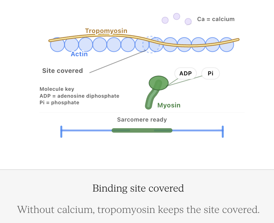
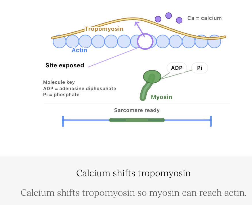
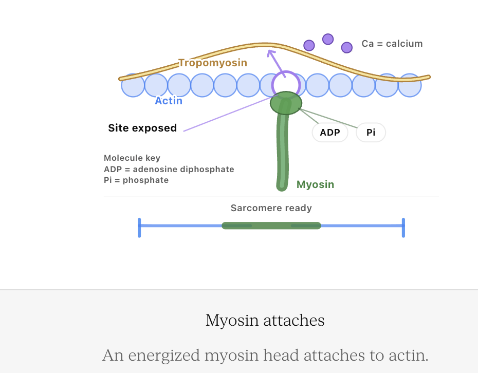
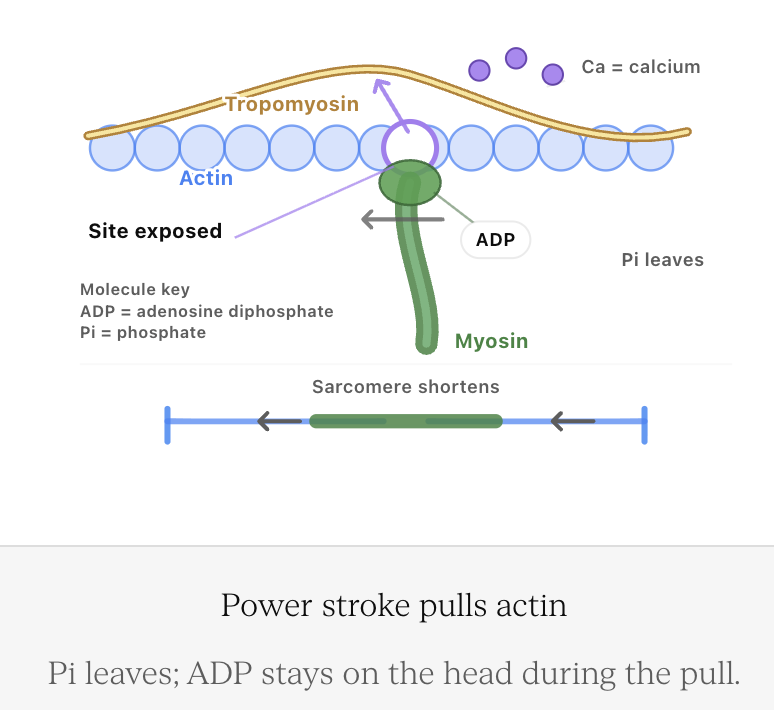
Actin and Myosin
Two important proteins involved in muscle contraction are:
- Actin
- Myosin
These proteins are arranged in repeating units called sarcomeres.
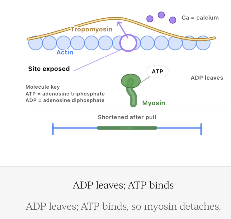
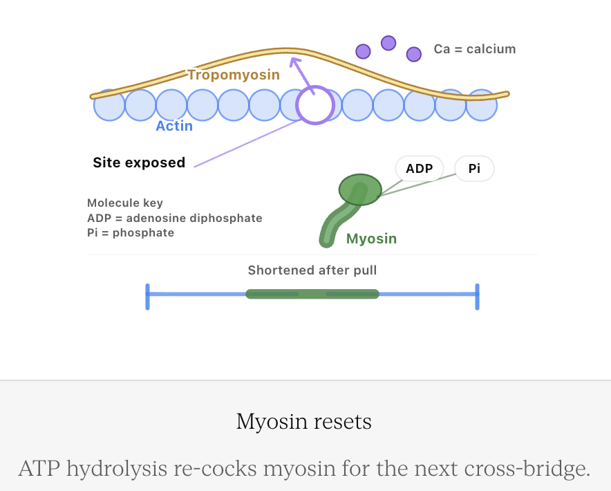
During contraction, actin and myosin interact so that the protein filaments slide past one another.
This is known as the sliding filament mechanism.


The filaments themselves do not simply become shorter.
Instead, their arrangement changes as they slide relative to one another, shortening the sarcomere.
Many sarcomeres shortening together causes the muscle fibre to shorten.
How Muscle Contraction Occurs
A skeletal muscle does not contract spontaneously during normal movement. It receives instructions from the nervous system.
A simplified sequence is:
Motor neuron stimulated
↓
Signal reaches muscle fibre
↓
Calcium ions become available inside the muscle fibre
↓
Actin and myosin interact
↓
Myosin pulls actin
↓
Sarcomeres shorten
↓
Muscle develops tension and may shorten


4. Skeletons and Support Systems
Learning outcomes
- I can compare hydrostatic, exoskeleton, and endoskeleton support systems.
- I can explain the functions of skeletons.
- I can describe how skeletons support movement and protection.
- I can identify adaptations of skeletal systems in different animals.
- I can explain the relationship between support and locomotion.
Why Do Animals Need Support Systems?
Animals need some form of structural support to maintain their shape and move effectively.
Without a support system, muscles would have little to pull against and many animals would be unable to maintain a stable body shape.
Support systems can perform several important functions:
- Support the body.
- Maintain body shape.
- Protect delicate organs.
- Provide attachment points for muscles.
- Allow movement.
- Support locomotion.
- In some animals, store minerals.
- Provide protection against predators or physical damage.
Animals have evolved several different solutions to the problem of support.
Three major types are:
- Hydrostatic skeletons
- Exoskeletons
- Endoskeletons


Each system has advantages and limitations and is suited to particular body forms and lifestyles.
What Is a Skeleton?
A skeleton is a structural support system that helps maintain body shape and allows forces produced by muscles to create controlled movement.
The word "skeleton" does not necessarily mean bones.
An earthworm, for example, has no bones but still has a support system that allows it to move effectively.
The three major systems work differently:
Hydrostatic skeleton → fluid provides support
Exoskeleton → rigid structure outside the body provides support
Endoskeleton → internal framework provides support
Hydrostatic Skeletons
A hydrostatic skeleton uses fluid contained within the body to provide support.
The fluid is largely incompressible, meaning that it does not easily decrease in volume when pressure is applied.
Muscles surrounding the fluid-filled space can change the animal's shape and produce movement.


Hydrostatic skeletons occur in animals such as:
- Earthworms.
- Many other worms.
- Sea anemones.
- Jellyfish.
Hydrostatic support also contributes to movement in structures such as the arms of octopuses.
How Does a Hydrostatic Skeleton Work?
Imagine a water-filled balloon.
The water inside cannot easily be compressed.
If one part of the balloon is squeezed, pressure is transferred through the fluid and the shape changes.
A hydrostatic skeleton uses a similar principle.
Muscles contract around a fluid-filled body cavity.
Because the fluid resists compression, muscular contractions can change the shape of the body and generate movement.
This allows the fluid to act as something for the muscles to push or pull against indirectly.
Earthworm Movement
Earthworms provide an excellent example of hydrostatic support.
They have two important sets of muscles:
- Circular muscles.
- Longitudinal muscles.
When circular muscles contract, a body segment becomes:
- Longer.
- Thinner.
When longitudinal muscles contract, the segment becomes:
- Shorter.
- Thicker.


Coordinated waves of contraction travel along the body.
Small bristles called setae help grip the ground.
The sequence can be simplified as:
Muscles contract → body shape changes → parts of body grip surface → animal moves forward
Advantages of Hydrostatic Skeletons
Hydrostatic skeletons have several advantages.
They are:
- Flexible.
- Lightweight.
- Capable of producing a wide range of body shapes.
- Useful for burrowing and squeezing through narrow spaces.
- Able to support soft-bodied animals without heavy rigid structures.
Flexibility can be particularly useful in aquatic environments and underground habitats.
Limitations of Hydrostatic Skeletons
Hydrostatic skeletons also have limitations.
They generally:
- Provide less rigid protection than hard skeletons.
- Depend on maintaining suitable internal fluid pressure.
- Provide fewer rigid attachment points for powerful muscles.
- May provide less support for large terrestrial animals.
For many small or soft-bodied animals, however, these limitations are not major disadvantages.
The effectiveness of a support system depends on the animal's environment and lifestyle.
Exoskeletons
An exoskeleton is a hard supporting structure located on the outside of the body.
Arthropods such as insects, spiders, crabs, and lobsters have exoskeletons.


Arthropod exoskeletons contain a material called chitin, often combined with other substances that change its strength and flexibility.
Functions of an Exoskeleton
An exoskeleton can perform several functions at once.
It can:
- Support the body.
- Maintain body shape.
- Protect internal organs.
- Provide attachment surfaces for muscles.
- Form joints for movement.
- Reduce water loss.
- Provide protection against predators and physical damage.
This combination has been highly successful. Arthropods are among the most diverse groups of animals on Earth.
Exoskeletons and Water Conservation
An important advantage of the arthropod exoskeleton is its ability to reduce water loss.
The outer surface can act as a barrier that limits evaporation.
This is especially important for terrestrial arthropods such as insects.
Without a protective outer covering, a small animal with a high surface-area-to-volume ratio could lose water rapidly.
The exoskeleton therefore contributes to both:
Support + water conservation
Exoskeletons and Movement
A rigid outer covering might seem as though it would prevent movement.
However, arthropod exoskeletons contain joints.
Flexible membranes between rigid sections allow body parts to move relative to one another.


Muscles attach to the inner surface of the exoskeleton.
When muscles contract, they move different sections of the exoskeleton around joints.
This allows movements such as:
- Walking.
- Running.
- Jumping.
- Swimming.
- Flying.
- Grasping.
A Major Problem: Growth
A rigid exoskeleton cannot simply expand continuously as an animal grows.
Arthropods therefore periodically shed their old exoskeleton.
This process is called molting, or ecdysis.


During a molt:
- A new exoskeleton develops beneath the old one.
- The old exoskeleton splits.
- The animal emerges.
- The new exoskeleton expands.
- The new covering eventually hardens.
The Risks of Molting
Molting allows growth, but it creates a period of vulnerability.
Immediately after leaving the old exoskeleton, the new exoskeleton may be relatively soft.
During this period, the animal may be:
- More vulnerable to predators.
- Less protected from physical damage.
- Less able to move normally.
- More vulnerable to water loss.
This is an important trade-off associated with having a rigid external skeleton.
Endoskeletons
An endoskeleton is an internal supporting framework.
Vertebrates have endoskeletons made primarily from:
- Bone.
- Cartilage.
Examples include:
- Fish.
- Amphibians.
- Reptiles.
- Birds.
- Mammals.


Some invertebrates, such as echinoderms, also have internal skeletal structures.
Functions of the Vertebrate Endoskeleton
The vertebrate skeleton performs several functions.
It:
- Supports the body.
- Maintains body shape.
- Protects organs.
- Provides attachment points for muscles.
- Allows movement around joints.
- Stores minerals.
- Contains bone marrow involved in blood-cell production.
The skeleton is therefore an active biological system, not simply a rigid framework.
Protection
Different parts of the vertebrate skeleton protect important organs.
For example:
Skull → protects brain
Vertebral column → surrounds and protects spinal cord
Rib cage → protects heart and lungs
Pelvis → helps protect organs in the lower abdomen and pelvic region


The shape of individual bones often reflects both their mechanical and protective functions.
Endoskeletons and Movement
Skeletal muscles are attached to bones by tendons.
When muscles contract, they pull on bones.
Bones then move around joints.
A simplified system is:
Muscle → tendon → bone → joint → movement
Bones can function as levers, while joints act as pivots.
This allows relatively small changes in muscle length to produce useful movements of limbs.
Antagonistic Muscles and the Skeleton
Because muscles pull rather than push, skeletal movement often requires antagonistic muscle pairs.
At the elbow:
Biceps contracts → forearm flexes
Triceps contracts → forearm extends


The endoskeleton therefore provides the rigid structures against which muscles can generate controlled forces.
Endoskeletons Grow with the Animal
Unlike an arthropod exoskeleton, a vertebrate endoskeleton can grow as the animal grows.
Bones are living tissues.
During growth, bone can:
- Increase in length.
- Increase in thickness.
- Change shape.
- Repair damage.
- Remodel in response to mechanical forces.
This means vertebrates do not need to shed their entire skeleton in order to grow.
Comparing the Three Support Systems
| Feature | Hydrostatic Skeleton | Exoskeleton | Endoskeleton |
|---|---|---|---|
| Main support | Pressurised fluid | Rigid external covering | Internal framework |
| Example | Earthworm | Insect | Human |
| Flexibility | High | Limited by rigid sections and joints | Moderate to high at joints |
| Protection | Usually limited | Strong external protection | Strong protection of selected organs |
| Muscle interaction | Muscles act against fluid | Muscles attach internally to exoskeleton | Muscles attach to bones |
| Growth | Can expand with body | Requires molting in arthropods | Grows with body |
| Water conservation | Usually limited | Can strongly reduce water loss | Skin provides main external barrier |
| Large body support | Usually limited on land | Size can be mechanically constrained | Can support large terrestrial animals |
No system is universally superior. Each represents a different solution to the problems of support, protection, and movement.
Support and Locomotion
Locomotion means movement of an animal from one place to another.
A support system makes locomotion more effective because it provides something against which muscles can generate forces.
The general principle is:
Muscle contraction + support system + interaction with environment → locomotion
For example:
- Earthworm muscles act against fluid.
- Insect muscles act against an exoskeleton.
- Human muscles pull on an endoskeleton.
Different structures, but the same fundamental requirement: muscular force must be translated into useful movement.
Support Systems and the Environment
The effectiveness of a support system depends partly on the environment.
Water provides buoyancy, which helps support an animal's weight.
This means aquatic animals may require less structural support against gravity than similarly sized terrestrial animals.


On land, gravity places greater demands on structures such as:
- Limbs.
- Vertebral columns.
- Joints.
- Muscles.
This helps explain why support structures differ between aquatic and terrestrial animals.
Adaptations for Running
Animals specialised for running often have skeletal adaptations that improve speed or efficiency.
These may include:
- Long limbs.
- Reduced mass toward the ends of limbs.
- Flexible spinal columns.
- Specialised joints.
- Elongated lower limb bones.


In a cheetah, for example, a flexible spine contributes to increased stride length during high-speed running.
Adaptations for Flying
Flight places very different demands on the skeleton.
Bird skeletons have several adaptations related to flight.
These can include:
- Lightweight bones.
- Fusion of some bones for strength.
- Modified forelimbs forming wings.
- Large surfaces for attachment of flight muscles.
- A streamlined body form.


Many flying birds possess a large keel on the sternum.
Powerful flight muscles attach to this structure.
The skeleton therefore provides both lightweight support and strong muscle attachment.
Adaptations for Swimming
Aquatic vertebrates also show specialised skeletal adaptations.
Fish typically have:
- Flexible vertebral columns.
- Fins supported by skeletal structures.
- Streamlined bodies.
- Muscles arranged to produce side-to-side movement.


The skeleton and muscles work together to push water backward.
The resulting forces move the fish forward.
Adaptations for Burrowing
Burrowing animals face yet another challenge.
They must move through soil or sediment.
Useful adaptations may include:
- Flexible bodies.
- Powerful digging limbs.
- Strong claws.
- Compact body shapes.
- Hydrostatic support in some worms.
An earthworm's hydrostatic skeleton is especially effective because the body can lengthen, shorten, and change shape while moving through narrow spaces.
Adaptations for Jumping
Animals specialised for jumping may have:
- Long hind limbs.
- Powerful muscles.
- Strong joints.
- Elastic structures that store and release energy.


Frogs, for example, possess elongated hind limbs that help generate large forces against the ground.
This demonstrates how skeletal proportions can be closely related to locomotion.
Protection Versus Mobility
Support systems often involve trade-offs.
A very thick, rigid skeleton might provide excellent protection but could:
- Increase body mass.
- Reduce flexibility.
- Require more energy to move.
A highly flexible support system might improve manoeuvrability but provide less physical protection.
Evolution therefore often produces compromises between:
Protection + support + flexibility + mass + energy cost
Different animals show different combinations depending on their environments and lifestyles.
Worked Example: Earthworm
Consider an earthworm moving through soil.
It has no rigid bones.
Circular muscles contract, causing sections of the body to lengthen.
Longitudinal muscles contract, causing sections to shorten.
Fluid inside the body resists compression.
Setae grip the soil.
The combination produces forward movement.
Therefore:
Muscles + fluid pressure + grip → locomotion
This is a hydrostatic skeleton in action.
Worked Example: Grasshopper
A grasshopper has an exoskeleton.
Its muscles attach to the inner surfaces of this external skeleton.
When leg muscles contract:
- Forces are transferred to rigid exoskeletal sections.
- Sections rotate around joints.
- The hind legs push strongly against the ground.
The ground exerts a force on the grasshopper, accelerating it upward and forward.


The exoskeleton therefore provides both support and a mechanical system for movement.
Worked Example: Human Arm
A human arm uses an endoskeleton.
When the biceps contracts:
- The muscle produces tension.
- The tendon transfers the force to the forearm.
- The forearm bones act as levers.
- The elbow acts as a pivot.
- The forearm moves.
Therefore:
Muscle contraction → tendon force → bone movement → movement at joint
The skeleton does not produce the force itself. The muscles provide the force, while the skeleton provides the mechanical framework.
Worked Example: Moving from Water to Land
Imagine an animal lineage moving from a mainly aquatic environment to a terrestrial environment.
In water, buoyancy supports much of the animal's weight.
On land, this support disappears.
Gravity places greater demands on:
- Limbs.
- Joints.
- Vertebral column.
- Muscles.
A successful terrestrial animal therefore requires structures capable of supporting its body against gravity while still permitting movement.
This illustrates how environment can influence the evolution of support systems.
Body Size and Support
As an animal becomes larger, its mass increases rapidly.
Its support system must withstand increasing forces caused by gravity and movement.
Large terrestrial vertebrates often have:
- Thick limb bones.
- Strong joints.
- Large muscle attachment areas.
- Limb positions that efficiently support body mass.


An elephant, for example, has thick, relatively column-like limbs that support its large mass.
A tiny insect can use a very different structural arrangement.
Skeletons Can Have Additional Functions
Support systems can perform functions beyond support and locomotion.
For example, vertebrate bones can:
- Store calcium and phosphate.
- Contain bone marrow.
- Produce blood cells.
- Protect organs.
- Store energy in yellow bone marrow.
An exoskeleton can also:
- Reduce water loss.
- Provide camouflage.
- Carry sensory structures.
- Provide defensive structures such as spines.
A skeleton is therefore often a multifunctional system.
Support Systems and Evolution
Hydrostatic skeletons, exoskeletons, and endoskeletons represent different evolutionary solutions to similar problems.
All must allow an animal to:
- Maintain body shape.
- Resist external forces.
- Produce controlled movement.
- Protect important structures.
- Function effectively in its environment.
The details differ because animals have different:
- Body sizes.
- Habitats.
- Feeding strategies.
- Predators.
- Methods of locomotion.
- Evolutionary histories.
Common Mistakes
Thinking Every Skeleton Is Made of Bone
Hydrostatic skeletons use fluid, and arthropod exoskeletons are largely composed of materials including chitin.
Thinking an Exoskeleton Is Just Protective Armour
An exoskeleton also provides support and attachment surfaces for muscles and plays an important role in movement.
Saying Exoskeleton Muscles Are Outside the Skeleton
In arthropods, muscles are located inside the exoskeleton and attach to its internal surfaces.
Saying Hydrostatic Skeletons Have No Structural Support
The pressurised fluid provides the resistance against which muscles act.
Saying Endoskeletons Do Not Protect the Body
Although they are internal, structures such as the skull, vertebral column, and rib cage provide substantial protection.
Thinking Skeletons Produce Movement by Themselves
Muscles produce force. Skeletons provide structures against which those forces act.
Saying Exoskeletons Cannot Move
Exoskeletons contain joints and flexible regions that allow movement.
Thinking an Exoskeleton Grows Continuously
In arthropods, the rigid exoskeleton must periodically be shed during molting.
Assuming One Skeleton Type Is Always Better
Each system has advantages and limitations. Its effectiveness depends on the animal's body form, size, environment, and lifestyle.
Check Your Understanding
1. Give four important functions of an animal skeleton.
2. Define a hydrostatic skeleton.
3. Explain how an earthworm uses its hydrostatic skeleton to move.
4. Give two advantages and two limitations of hydrostatic skeletons.
5. Define an exoskeleton and name two groups of animals that possess one.
6. Explain how muscles produce movement in an animal with an exoskeleton.
7. Why must arthropods molt?
8. Explain one disadvantage of molting.
9. Define an endoskeleton.
10. Explain how an endoskeleton works with muscles to produce movement.
11. Compare the growth of an exoskeleton with the growth of a vertebrate endoskeleton.
12. Explain why an exoskeleton can be useful for a small terrestrial animal.
13. Describe two skeletal adaptations for flight, running, swimming, or jumping.
14. Explain why large terrestrial animals require particularly strong support structures.
15. An unknown animal has a soft flexible body, no rigid skeleton, and muscles surrounding a fluid-filled cavity. Identify its type of support system and explain how it could use that system for locomotion.
Key Terms
- Skeleton – structural system that supports an animal and helps muscles produce controlled movement.
- Support system – structures or mechanisms that maintain body shape and resist forces.
- Hydrostatic skeleton – support system in which muscles act against pressurised, relatively incompressible fluid.
- Exoskeleton – rigid supporting structure located outside the body.
- Endoskeleton – internal supporting framework.
- Chitin – structural material forming an important part of arthropod exoskeletons.
- Molting – shedding of an old exoskeleton to allow growth.
- Ecdysis – another term for molting.
- Bone – strong living connective tissue forming much of the vertebrate skeleton.
- Cartilage – flexible supportive connective tissue.
- Joint – location where skeletal structures meet and, in many cases, permit movement.
- Tendon – connective tissue attaching muscle to skeletal structures.
- Lever – rigid structure that rotates around a pivot when a force is applied.
- Locomotion – movement of an organism from one location to another.
- Buoyancy – upward force exerted by a fluid that helps support objects immersed in it.
- Setae – small bristles that help organisms such as earthworms grip surfaces.
- Antagonistic muscles – muscles that produce opposing movements.
- Keel – projection of the sternum in many birds that provides attachment for major flight muscles.
Key Takeaways
- Animals require support systems to maintain shape, protect organs, and produce effective movement.
- The three major support systems are hydrostatic skeletons, exoskeletons, and endoskeletons.
- Hydrostatic skeletons use pressurised fluid as a supporting structure.
- Earthworms use circular and longitudinal muscles acting against fluid to produce locomotion.
- Hydrostatic skeletons provide excellent flexibility but relatively little rigid protection.
- Exoskeletons form rigid structures around the outside of the body.
- Arthropod exoskeletons provide support, protection, muscle attachment, and reduced water loss.
- Joints allow animals with exoskeletons to move despite their rigid outer covering.
- Arthropods must periodically molt because their rigid exoskeleton cannot continuously expand.
- Endoskeletons are internal support structures that can grow with the animal.
- Vertebrate endoskeletons consist largely of bone and cartilage.
- Bones protect important organs and provide attachment points for muscles.
- Muscles produce force while skeletons provide the structures against which those forces act.
- Bones and rigid exoskeletal sections can function as levers during movement.
- Aquatic animals benefit from buoyancy, while terrestrial animals require greater structural support against gravity.
- Skeletal adaptations can support specialised forms of locomotion such as running, flying, swimming, jumping, and burrowing.
- Support systems involve trade-offs between strength, protection, flexibility, mass, and energy cost.
- The structure of an animal's support system is closely related to its size, environment, lifestyle, and method of locomotion.
5. Coordination of Responses
Learning outcomes
- I can explain how animals coordinate responses to stimuli.
- I can describe the pathway of a simple reflex action.
- I can explain how different organ systems work together during responses.
- I can distinguish between voluntary and involuntary responses.
- I can analyze how coordinated responses improve survival.


