Cell Division and Growth

Site: Young Education
Cours: Cells and Life Processes
Livre: Cell Division and Growth
Imprimé par: Gwestai
Date: lundi 5 octobre 2026, 05:00

1. The Cell Cycle

Learning outcomes
  • I can describe the stages of the cell cycle.
  • I can explain the purpose of the cell cycle in living organisms.
  • I can identify the events that occur before, during, and after cell division.
  • I can explain why DNA must be replicated before cell division.
  • I can relate the cell cycle to growth and repair.

The Cell Cycle

Living organisms grow, replace damaged cells, and maintain tissues by producing new cells. New cells do not simply appear. They are produced when existing cells go through an organized series of events called the cell cycle.

During the cell cycle, a cell:

  • grows
  • carries out its normal functions
  • copies its DNA
  • prepares for division
  • divides its nucleus
  • divides into two new cells

The two cells produced are called daughter cells.

https://images.openai.com/static-rsc-4/Yu353kTO2WCWMsDu3uN1XrfXe2YCaNyLNDywGtn8hRQRMhb4O22dM7j24Rp0hf3sJVFdU8CnBn0L_zrs1yJb9N6lOEUb3KVuFinOJNPg1kxlU643CvRaI8XDaAwCJFcklEiHwEWFNWkcHpeYTJ19i1TJvh41AJXN6CEZQwT-8uvYqHza-UNgKw0H3PaMRl_0?purpose=fullsize
 
https://images.openai.com/static-rsc-4/n9y_bO5fc0998_XeJeFsZ2ridWlhh6Ghrqf_KhaYAYVtuH96Z09WUwdOzzT1bEJ_FLGxqgVDIRT8xOA0ou3CS7m36zcVI_ECv9BKU9S5BOPK5j1jrFmkGALxUhzgHpm763iHvvvzLA1l3yS_XSPHR5FIM9ZCOmzbfaBX-wUyoz6jfB5AJv1YEixldPce75f0?purpose=fullsize
 
https://images.openai.com/static-rsc-4/cml_xko85XZgpXUSlyTLY7fQFf9AQkSh_1H4lSJAs64x94Cd200T0o5DabEKgbBCY0CtSsB_b8j9FkwfPGr5PJZiiIL_qxgQhq0TtUdfjYLiojYmzTaykIy34N6h7DlSRcngSqfbTCo_hMubCxX9ObnNe8E2V7M8L8t-wGWKw2nXoeSPk-E7AF_vyIfaY7qt?purpose=fullsize
 
5

What Is the Cell Cycle?

The cell cycle is the series of stages that a cell passes through as it grows and divides.

A simplified cell cycle can be divided into three major parts:

Interphase → Mitosis → Cytokinesis

During interphase, the cell grows, performs its normal activities, copies its DNA, and prepares for division.

During mitosis, the copied chromosomes are separated into two nuclei.

During cytokinesis, the cytoplasm and cell membrane divide, producing two daughter cells.

The cycle can then begin again.


Why Do Cells Divide?

Cell division is necessary for several important biological processes.

These include:

  • growth
  • development
  • repair
  • replacement of old cells
  • replacement of damaged cells
  • asexual reproduction in some organisms

A multicellular organism begins as a single cell.

Repeated cell division increases the number of cells:

1 → 2 → 4 → 8 → 16 → 32 → ...

Eventually, millions or even trillions of cells can form.


The Main Stages of the Cell Cycle

The cell cycle can be summarized as:

Before cell division

Interphase

The cell grows, carries out normal activities, replicates its DNA, and prepares for division.

During nuclear division

Mitosis

The chromosomes are separated so that two nuclei form.

After nuclear division

Cytokinesis

The cytoplasm divides and two daughter cells are produced.

This process can be followed visually below.
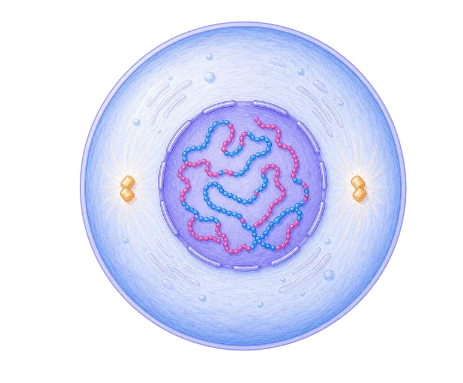
Interphase
 
DNA has been copied before mitosis, but it still remains uncondensed inside one nucleus.
 
 
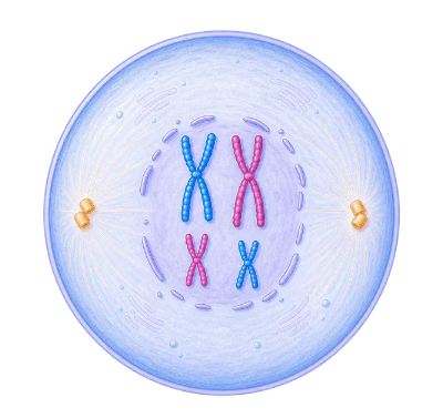
Prophase
Chromosomes condense and the spindle starts to form.
 
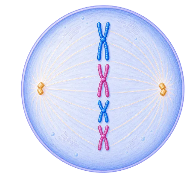
Metaphase
Duplicated chromosomes line up across the middle.
 
 
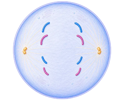
Anaphase
Sister chromatids move toward opposite sides.
 
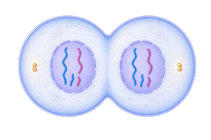
Telophase + Cytokinesis
Two identical daughter cells finish forming as new nuclei reappear around decondensing DNA.
 

Interphase

Cells spend much of the cell cycle in interphase.

Interphase is sometimes incorrectly described as a "resting stage."

The cell is not resting.

During interphase, it is extremely active.

The cell may:

  • grow
  • produce proteins
  • make new organelles
  • carry out its normal functions
  • replicate its DNA
  • prepare for division
https://images.openai.com/static-rsc-4/JX84EOYLu-SiLBkF5zXhCsUpd7UJq-sjRWYGj3BBWKcfRq2YqL6CeF28QNXMoQBXific-VSYxxlizJ7IeyZFwYx3UfrEhqd-go45Q6ZPEBFYVW_GJUqjfIiDmyqQMAKLwZKIOlQ7P03oxV9GBZd_vLg7dVGYE7iuZfn3sFK8xRSl17jWoA1d3CIQucPgwCoV?purpose=fullsize
 
https://images.openai.com/static-rsc-4/HLjAiYcXXa0slVzTbHMpkol_o9q5R2ibpud2TSEpDT9Qt5rOfI6k_3X7LolKnJxJIPsiluiRO6s-aKrAoJoflI8rFRHctKD9jAgI3bnM0JUVZ6FdwJqJaoRuJ78tVn38iBxtaBX--Devna39vSkyaoSwV0upQIKICJn8RoH8pttqA9BnMaOy3_0e5ia9CFxI?purpose=fullsize
 
https://images.openai.com/static-rsc-4/fO8eGCdpns6NaYRUXUW6SaZ8L2KoO-q8Uf__3-DGWbd9mb0SgBeRHg0XaW0ub5NF3UUu9FGpyHJP3Oet_X7Hbo2C3ddfEPKZFB5A_OjQukuLkEpdFo9umwuOKqmUh4gCYyYUD9Z06q1teV4oMCCWL-o_BAImhro3r962xVgxhtZeU1BfZZqu2HsaeyE5Bm3E?purpose=fullsize
 
6

The Three Parts of Interphase

Interphase can be divided into three stages:

G1 → S → G2

G1 Phase

During G1, the cell:

  • grows
  • carries out normal functions
  • produces proteins
  • increases the number of some organelles

The letter G stands for gap, although this is a period of considerable cell activity.

S Phase

During the S phase, the cell replicates its DNA.

S stands for synthesis.

Each chromosome is copied so that there will be enough genetic information for both daughter cells.

G2 Phase

During G2, the cell:

  • continues growing
  • produces molecules needed for division
  • checks and prepares its DNA
  • prepares the structures required for mitosis

After G2, the cell can enter mitosis.


DNA Replication

Before a cell divides, its DNA must be copied.

This process is called DNA replication.

DNA contains the genetic instructions needed for the cell to function.

If a cell divided without copying its DNA first, the daughter cells would not receive complete sets of genetic information.

Therefore:

DNA replication must occur before mitosis.

https://images.openai.com/static-rsc-4/bSED3kdrJ6PDvqKvmFst_ehF7_YM4_CERA_4xR7K0D2QyN3yFRlqPiPlb59ANwx9v6Su6FAqEyft2ZemCiIzaBNpNCQPWerN3HUlFHEHbuMr2Fo3w5kaWiorPvD-st1ZXmfDpq74oQ5j_PZTWX1sL7St5uS4aKx755MNLuUcSKmD4nn0gXo00XltONPhtSGH?purpose=fullsize
 
https://images.openai.com/static-rsc-4/mnhtNZFgxtVny0sb8gpi0lyvKmZvi8asl6VucUw6G_PeTtEhHvM50NcPOpLPlTCc8RCalH-2o35cgmKVUaeBS7CSfNQBdVCeb5A_pe6tdBHPZO_9U0A5PrYMZYwu03Uy7Nz83IlX01rDHhvYXj2IPuPhzIguSn4jPzikT4x1iE_jlMEU9hWBcN7IZQsGJs9v?purpose=fullsize
 
https://images.openai.com/static-rsc-4/JxYB3Ur6ia6dd6BoPvPIaNDANGPylFKD5NxuU5DpfvgxnAW2M5WCGP59Y8oT35YXmjsDrA1fwOimKMLnCQPQxWDeFRj-YiuxgJOBPV3BDDMqTk42HeAb98CMSIzLLHp1xmFfPbXcfB0YVLRiBM0QZYJ1DfYlb0veOPdyYY36GGmJHtAvU6un2Kn3DtqbD_BW?purpose=fullsize
 
6

Why Must DNA Be Replicated?

Imagine a cell contains one complete set of genetic instructions.

If it simply divided those instructions between two daughter cells without copying them, each cell would receive only part of the information.

Instead, the cell first produces a copy.

This creates two complete sets of DNA.

During mitosis, the copied chromosomes are separated.

As a result, each daughter cell receives a complete set of genetic information.


Chromosomes Before Cell Division

DNA is organized into structures called chromosomes.

Before DNA replication, each chromosome consists of one DNA molecule.

After replication, the chromosome consists of two identical copies called sister chromatids.

The sister chromatids are joined at a region called the centromere.

A replicated chromosome is often drawn as an X-shaped structure.

However, chromosomes do not remain X-shaped throughout the entire cell cycle. This shape becomes particularly visible when chromosomes condense during cell division.


Mitosis

Mitosis is the division of the nucleus that produces two genetically identical nuclei.

Mitosis ensures that each daughter cell receives the same number and types of chromosomes as the original cell.

Mitosis is commonly divided into four main stages:

Prophase → Metaphase → Anaphase → Telophase

A useful memory aid is:

P → M → A → T

https://images.openai.com/static-rsc-4/hym4cEEF4TPSXRJqFhXpNtIzawT_8H_DNdkbhN162Zx4Ex8iGk1wzRfHzZmYG7eiRGn3G6hIwLLjK6AQPc8JMgXZQRK2GG2tiQwiViY-FS0BlCVG1Wy0Iy-bxJrctmM7cmHzCQVS87tZDHUGZH40jVYU3jxAMNtebs4o7n45jaMk8SYn4t0Fe49Rald2r5vV?purpose=fullsize
 
https://images.openai.com/static-rsc-4/sdS35Lg8JBb_C-Ei9NgiKBV_FDH4W-oFzntUtlI9acgwx5-nYeQtqwKvcF9BBpblhazd1ucamrO5Xb4S61yiw7cGO11jZHK35xzGv25_m4BvrfVsGhdT4ZwwPNmUa8gEDqBO7iD50ol0RUoSlqA4rqgUfTCFYAbluBXjlEAEhrFHpUbJ5CJDIoPNGOqG_5ZB?purpose=fullsize
 
https://images.openai.com/static-rsc-4/No83DDUXq8RbAibEuwnuzl-g7eIpyGYOrdtAyt6KMun_f-RFLpdQacMGbrkyoAFEIXVVWrL62SlnOWd-Dafvh3s6deYCRTDabgATVqFnMeP1xJfTk6b3gI8pdUwumkn3J24z1LouWBQKkBI7MLcMJSTm_uioXsIzexa8tVp86E0ACR4E132I-IPejQ7SFPRM?purpose=fullsize
 
5

Prophase

During prophase:

  • chromosomes condense and become more visible
  • each replicated chromosome consists of two sister chromatids
  • the nuclear envelope begins to break down
  • spindle fibres begin to form

The chromosomes become compact, making them easier to move without becoming tangled.


Metaphase

During metaphase:

  • chromosomes move toward the middle of the cell
  • chromosomes line up around the cell's equator
  • spindle fibres attach to the chromosomes

The alignment helps ensure that the chromosome copies can be separated accurately.

A useful clue for identifying metaphase is:

chromosomes lined up across the middle


Anaphase

During anaphase:

  • sister chromatids separate
  • spindle fibres pull them toward opposite ends of the cell

Once the sister chromatids separate, each becomes an individual chromosome.

A useful clue for identifying anaphase is:

chromosomes moving apart


Telophase

During telophase:

  • chromosomes reach opposite ends of the cell
  • chromosomes begin to uncoil
  • new nuclear envelopes form
  • two nuclei are produced

Mitosis is now nearly complete.

The cell has divided its genetic material into two nuclei.


Identifying Mitosis Under a Microscope

Scientists can observe dividing cells using microscopes.

Growing regions of plants, such as onion root tips, are particularly useful because many cells are actively dividing.

https://images.openai.com/static-rsc-4/Hfw5FPk6VKym3nPgrF985MQZD4AvXMcjCD0KWrWbGpl4p_EPfjf5PUMN9Rnfu9nnthsfz26XVK122_3Wqx1jya8U8K3lNbtg4MjYgX2zCmOStsKb1tX-xq_9S5mPAzHkspoJXPTQWq1gFweDXVU0a297aC6JL2Z8krabMXKR3eRlt3EnRSPBZxb7m3RaIqd3?purpose=fullsize
 
https://images.openai.com/static-rsc-4/gw8pgYAAwugW2b6tUrvZLzmlq96DFMM0AivSOKYcot_f8jhpvYu613kVHdN7O3xt401cViG2ps-Ur8ZAO3WkpDwJXa2udsgEjAcAqNUXWWxDC73RJFiilvlNhytc519iVJxaht3-mRyCMm9_8KmN0StcLpUTjzNZNNOqlTRmDZoOqWuI7QitCk__pFSUvUxv?purpose=fullsize
 
https://images.openai.com/static-rsc-4/peY4Q2lyhA14eD0BMrokeap8YtHAIUHkh14z3qC9mdgUYUkQ8bFdzM1RY8BWIvx89v2Fi4HpSJ_M_tIsD0iZ3UgOrfckbfyFyfvmHQkX0XIo5Pwn1Dq__AAItz8BL9G6mFtYUS7s3PIVqEpTiA54aEs2quD71bicEDL0tllQCHImY_20PE7sGIZzb1ZMGbSZ?purpose=fullsize
 
5

When examining cells, look for:

  • visible condensed chromosomes
  • chromosomes lined up in the centre
  • chromosomes separating
  • two nuclei beginning to form

Many cells will appear to be in interphase because cells generally spend much more time in interphase than in mitosis.


Cytokinesis

After the nucleus has divided, the cell itself must divide.

This process is called cytokinesis.

During cytokinesis:

  • the cytoplasm divides
  • the cell separates
  • two daughter cells form

Cytokinesis occurs differently in animal and plant cells.


Cytokinesis in Animal Cells

Animal cells do not have rigid cell walls.

During cytokinesis, the cell membrane pinches inward.

A groove forms around the middle of the cell.

This is called a cleavage furrow.

The membrane continues pinching until two separate daughter cells form.

https://images.openai.com/static-rsc-4/dxRc1mUOPWWSqew8DOX436GgKYnVLTxBEuiaAa5M5ac8UVJkcRFQRS8r1Oaf6i5ziLRCatPGN9lb1s2ruWOS5kNAbsrr2-aaSaMRp4EGoi30tPS95Y9V3l_QxJm35SJVaNQuMufJMOF3GI3THnFA2ClqCqDa3cEdnaNJypaOvW4ZY2aL5wZ7gbOLKjBqknOp?purpose=fullsize
 
https://images.openai.com/static-rsc-4/35phO7X-C0yAF_Ew8tO8xvrXJoyg1fQNnosDZcfTPLlGiNyp2xZ5aUQpAQLiIMj-3qO3Cw-LyhQP4XXU-GOurbxtsDxQwX0QcBqyRkzkms5s8c749yJiNkHs2YIspwreK6aNs3c8CT04Es2sup_LuRPE1dfMLUbI7UWpqm6F2EqPFXPCsWQ0h2FqdBWhO5Xp?purpose=fullsize
 
https://images.openai.com/static-rsc-4/Hf-2rM0Aub5Rqep3zfHQIT3EwTpuENE9463Un6oamI4Rnb-vW9aKWnIRM69QJmu-JAW4peYg5bzPAS7-7WQrHYIAcjD0w6zCebmXJxw4LKwQmBH1q5i5ub6nOCRReBeObkF5BpVP3CMNbwYRrUvDAaBktPGeOpWbKyzlzZ_Tv1OZfgwBbpAZIqbQujdo2t5l?purpose=fullsize
 
4

Cytokinesis in Plant Cells

Plant cells have rigid cell walls, so they cannot simply pinch inward.

Instead, a structure called a cell plate forms between the two new nuclei.

The cell plate develops into new membranes and a new cell wall.

This separates the original cell into two daughter cells.

Therefore:

Animal cells → cleavage furrow

Plant cells → cell plate


The Complete Cell Cycle

The overall sequence is:

G1 → S → G2 → Mitosis → Cytokinesis

The major events are:

Stage Major Event
G1 Cell grows and performs normal functions
S DNA is replicated
G2 Cell prepares for division
Prophase Chromosomes condense
Metaphase   Chromosomes line up
Anaphase Sister chromatids separate
Telophase Two nuclei form
Cytokinesis Cell divides into two

What Are Daughter Cells?

The cells produced by cell division are called daughter cells.

For normal mitotic division, the daughter cells are genetically very similar to each other and to the parent cell.

They receive:

  • a complete set of chromosomes
  • cytoplasm
  • cell membranes
  • cellular structures

After division, the daughter cells may grow and eventually enter another cell cycle.


The Cell Cycle and Growth

Multicellular organisms grow mainly by increasing their number of cells, rather than simply making individual cells larger and larger.

Imagine a fertilized egg.

It begins as one cell.

That cell divides:

1 cell → 2 cells

The cells divide again:

2 → 4

Then:

4 → 8

Repeated cell cycles eventually produce the enormous number of cells making up an organism.

https://images.openai.com/static-rsc-4/8s2Ltek7Q3j-ao3ZaIHyNgJ1vv-hc2fBgUEDCAsDMfG8l8QXc6FSJNEsS-85PnLXjA4Z6xdZgmcAEnmfrz811zG-UaEO5VlrwWq8KKuhJ0lAUseBMIk3gKo0Su1YWc0DcU8ry3e7zdgv5-vUQ4-TmqoMLJKATFCm7SYnSQTCK2kS79t3sH-RfkcMfcnWIITQ?purpose=fullsize
 
https://images.openai.com/static-rsc-4/azQ2tOBL8HeFu4LLFuSTxYpI_PCL8gWG6-GkNlmoL1YwlfkpQn94D_9H1CqHBoXLAdDaQ-ji_aC7vMN87ncQgM8Fe5RFyCMDrLmVfJPPQ7ZS9ild5qliS74U2rns8aq2ZHMh-kbyLhYoxnvi2qDf5IUtu05gMWgnSbcYQL_GCxVydoeEhsrlbDqtvBQirL-l?purpose=fullsize
 
https://images.openai.com/static-rsc-4/KnkFc8xQ4_9FaizXBPwox6VSGcMIsOvnH8Ej8Oua_-vACg7sml8GBED0udhF6InVlXiRrG83fb2Pp7u6lao5088zVhEg-ohjJDm5-LceVk3dTOZOrIIFc65TeL6gOSrvd4YhMc4I8H9am1BQC0QOhQRARlqfAWfJfkgbiFJEZTBh85jgYlDzl4DWvQZBVH5y?purpose=fullsize
 
5

Cell division therefore plays a major role in:

  • embryonic development
  • childhood growth
  • tissue development
  • growth throughout an organism's life

The Cell Cycle and Repair

Cells can become damaged through:

  • injury
  • friction
  • chemicals
  • heat
  • radiation
  • normal wear and tear

Damaged or lost cells often need to be replaced.

Cells near the damaged area can undergo the cell cycle and produce new cells.

For example, if you cut your skin, cells around the wound divide and help replace damaged tissue.

https://images.openai.com/static-rsc-4/jPq4V_NtRIhFZIlh4V8zmT-mTbGI0iMpSy1ITqQo9xCbcvJWhi0SA9R-q2zRAQudlpfz79hwRmgDAgkK54oBXYkRxrBKx-aRfu1uI0glpc3VaQn4-d9U-_JQuTs2nKv1-rmfVvJ_4tVIPVdunEHGoE66WywL0T2MM8la-MJzTBjFAWlqTyA9B4mCB85QmPLY?purpose=fullsize
 
https://images.openai.com/static-rsc-4/6si-PKZE7OqrauPkOVXZN5amvyodpTanK3xjnwAxV_ugci3kE-7ru5tx5qrTaXfpYTxKfA53qQ-gaZikHbK5ruXZXp1mbuh7WbZiF3GBqf_mLpCosFhWpNBvdqQUNww8mGNZRYQ2o2DD2BLQiiSlcaiXW8-bc5dxOY8u1KVL8doisbcQ5hHHeJVNBdWkH9f9?purpose=fullsize
 
https://images.openai.com/static-rsc-4/To2vzjiykgGqWWB1qdsTrE7yBqM_6dZlmsOu2ftS0MuRkW3RzqNN9DLC-P8thqLq6T-ZsBRPvRTVmuBPszC2K6skiwKefJ3Bsfz-A9TOoeJo0hP-_wKaIP9HoKTLG8ol4s6QlL9v020lIAZBaUloF7bdrWgWtympIBDHs8vEeYloKuod5hGycI1m8u133Daj?purpose=fullsize
 
5

This is why cell division is essential for tissue repair.


Replacing Old Cells

Many cells do not survive for the entire lifetime of an organism.

They must constantly be replaced.

Examples include cells associated with:

  • skin
  • digestive tract lining
  • blood production

Cells in different tissues divide at different rates.

Some tissues undergo frequent cell division because cells are regularly lost or damaged.

Other specialized cells divide much less frequently.


Cell Division and Surface Area

Cell division also helps cells remain small.

As a cell grows, its volume increases faster than its surface area.

This reduces its surface area-to-volume ratio.

Eventually, exchange across the membrane may become less efficient relative to the needs of the cell.

Cell division produces two smaller cells.

These smaller cells have higher surface area-to-volume ratios, making exchange more efficient.

This connects the cell cycle to the concept of surface area and exchange.


Controlling the Cell Cycle

Cells do not normally divide continuously without control.

The cell cycle contains checkpoints.

At these checkpoints, the cell can assess whether conditions are suitable for continuing.

Checks may include:

  • Is the cell large enough?
  • Has the DNA been copied correctly?
  • Is the DNA damaged?
  • Are the chromosomes correctly attached before separation?

If serious problems are detected, the cell cycle may stop.

This helps prevent damaged cells from dividing.


What Happens When Cell-Cycle Control Fails?

Normally, cell division is carefully controlled.

Sometimes mutations affect genes involved in controlling the cell cycle.

A cell may then begin dividing when it should not.

Repeated uncontrolled cell division can produce a mass of cells called a tumour.

Some tumours can develop into cancer.

This is one reason why regulation of the cell cycle is extremely important.


Worked Example: Why Copy DNA?

A cell is preparing to divide into two daughter cells.

Why must it replicate its DNA first?

The cell needs to produce two complete copies of its genetic information.

During mitosis, one complete set can then be distributed to each new nucleus.

Therefore:

DNA replication ensures that each daughter cell receives a complete set of genetic information.


Worked Example: Identifying a Stage

A microscope image shows chromosomes lined up across the middle of a cell.

Which stage is occurring?

The key clue is:

chromosomes lined up in the middle

Therefore, the cell is in:

metaphase


Worked Example: Identifying Anaphase

A student observes two groups of chromosomes moving toward opposite ends of a cell.

This indicates:

anaphase

because sister chromatids have separated and are being pulled toward opposite poles.


Worked Example: Growth

A student says:

"A child grows mainly because every cell becomes much larger."

This is incomplete.

Although cells can grow, multicellular organisms increase in size largely by producing more cells through cell division.

Therefore, repeated cell cycles contribute directly to growth.


Worked Example: Repair

A person scratches their skin and damages some surface cells.

Cells near the damaged area enter the cell cycle.

They:

  • grow
  • replicate their DNA
  • undergo mitosis
  • complete cytokinesis

The new cells replace damaged cells.

Therefore, the cell cycle contributes to tissue repair.


Before, During and After Cell Division

A useful way to organize the cell cycle is:

Before division

The cell:

  • grows
  • carries out normal functions
  • replicates DNA
  • prepares for division

During nuclear division

The cell:

  • condenses chromosomes
  • lines them up
  • separates chromosome copies
  • forms two nuclei

After nuclear division

The cell:

  • divides its cytoplasm
  • forms two daughter cells
  • begins growth again

Mitosis Is Not the Entire Cell Cycle

One common mistake is using mitosis and cell cycle as if they mean the same thing.

They do not.

The cell cycle includes:

Interphase + Mitosis + Cytokinesis

Mitosis is only the stage in which the nucleus divides.

In fact, cells generally spend much more time in interphase than they do undergoing mitosis.


Common Misconceptions

Interphase is a resting stage.

Incorrect. Cells are highly active during interphase. They grow, perform normal functions and replicate their DNA.

Mitosis is the entire cell cycle.

Incorrect. Mitosis is only one part of the cell cycle.

DNA is copied during mitosis.

DNA is replicated before mitosis, during the S phase of interphase.

Chromosomes are always X-shaped.

No. The familiar X shape represents a replicated and condensed chromosome consisting of two sister chromatids.

Cell division is only needed for growth.

It is also important for tissue repair, cell replacement and asexual reproduction in some organisms.

Mitosis produces four cells.

Normal mitotic cell division produces two daughter cells.

Plant and animal cells complete cytokinesis in exactly the same way.

Animal cells form a cleavage furrow, while plant cells form a cell plate.


Did You Know?

Different cells in your body move through the cell cycle at very different rates.

Cells in tissues that experience frequent wear, such as the lining of the digestive system, must be replaced regularly.

Other highly specialized cells may remain outside the active cell cycle for very long periods.

This means cell division is carefully matched to the needs of each tissue.

https://images.openai.com/static-rsc-4/madAkY7gM398fJcLiOBEJhmwY3SYdLHwx-o-ulUYkBDTrdruKD0jDf_mmbOsa5hgLKH5EfD3CnPBZJlN8BDyhVyTWTF0qO1BeWsuyB6xs8prhn9ahbADy4G909d_q7erxX8Mr4us1gqy6xtlCkZkkVdJVPuAuJFxkMTiskvITGASonApGHUsMga779g3TtVh?purpose=fullsize
 
https://images.openai.com/static-rsc-4/Whi0SGr2ONEuq-imJO3JTLw2uVDzvGdHqqSEhpYU7Se7QgOYO-CwPJkCHYGBJKncIMUfza8kgrAZ0t8gLp3c7jL7h0oun3eikLHTU7pmd-8Zd657Wnz54rr1zuL1yorlECb8nscczL0VJ-j1Szyo0bMZBYPS_WwNnb8jk-yCSlEpeVFg6k7EwVMCsECXIkAH?purpose=fullsize
 
https://images.openai.com/static-rsc-4/4WwcHLxSQS3bG93Keg2IwnlEWrM1N0ZnwUs1ypB-2SGWUce70rw4BT6-9pIBm0zuG8bf3ZdiulDmLdYJp3Da7UD-CyqmOsAmLE0dejU3-Oa9vUxMU9aK8e06uTsZD1cl06uHzppSCgjd2i4H4m3fzQslYlgNy1567uE362VqsJGEdc0usmIxdf-XHhl0-MLJ?purpose=fullsize
 
6

Key Terms

Cell cycle – The series of stages through which a cell grows, copies its DNA and divides.

Interphase – The part of the cell cycle during which the cell grows, functions, replicates its DNA and prepares for division.

G1 phase – A stage of interphase involving cell growth and normal activity.

S phase – The stage during which DNA replication occurs.

G2 phase – The stage during which the cell prepares for division.

DNA replication – The process of copying DNA before cell division.

Chromosome – A structure containing DNA and associated proteins.

Sister chromatids – Identical copies of a replicated chromosome.

Centromere – The region where sister chromatids are joined.

Mitosis – Division of the nucleus to produce two genetically similar nuclei.

Cytokinesis – Division of the cytoplasm to produce two daughter cells.

Daughter cells – Cells produced by cell division.

Cell plate – A structure that forms between new plant cells during cytokinesis.

Cleavage furrow – The inward pinching of an animal cell membrane during cytokinesis.

Key Takeaways

  • The cell cycle is the sequence of events through which cells grow and divide.
  • The main parts are interphase, mitosis and cytokinesis.
  • During G1, the cell grows and carries out normal functions.
  • During the S phase, DNA is replicated.
  • During G2, the cell prepares for division.
  • DNA must be copied before division so each daughter cell receives a complete set of genetic information.
  • Mitosis can be divided into prophase, metaphase, anaphase and telophase.
  • During mitosis, replicated chromosomes are separated into two nuclei.
  • During cytokinesis, the cytoplasm divides.
  • Mitotic cell division normally produces two genetically similar daughter cells.
  • Animal cells divide using a cleavage furrow, while plant cells form a cell plate.
  • The cell cycle allows multicellular organisms to grow by increasing their number of cells.
  • Cell division also replaces old or damaged cells and allows tissue repair.
  • Cell-cycle checkpoints help ensure that cells divide correctly.
  • Loss of normal cell-cycle control can lead to uncontrolled cell division and tumour formation.


2. Mitosis

Learning outcomes
  • I can define mitosis.
  • I can describe the stages of mitosis.
  • I can explain how mitosis produces genetically identical cells.
  • I can identify the role of chromosomes during mitosis.
  • I can explain the importance of mitosis for growth and tissue repair.

Mitosis

Mitosis is the process in which the nucleus of a cell divides to produce two genetically identical nuclei.

It is an essential part of the cell cycle and allows organisms to:

  • grow
  • replace worn-out cells
  • repair damaged tissues
  • reproduce asexually in some organisms

Mitosis ensures that each new cell receives the same genetic information as the original parent cell.

https://images.openai.com/static-rsc-4/hym4cEEF4TPSXRJqFhXpNtIzawT_8H_DNdkbhN162Zx4Ex8iGk1wzRfHzZmYG7eiRGn3G6hIwLLjK6AQPc8JMgXZQRK2GG2tiQwiViY-FS0BlCVG1Wy0Iy-bxJrctmM7cmHzCQVS87tZDHUGZH40jVYU3jxAMNtebs4o7n45jaMk8SYn4t0Fe49Rald2r5vV?purpose=fullsize
 
https://images.openai.com/static-rsc-4/_XIE96AUYSOP-y3mCVHSNJJ3sZN6yqqTo2Xty8bWjiz9h1WqGIgvG7bSAP8zW5O1b86I8jql8r3fQLJT3oqBGzUR-sV6VH0az4of41vXGwhvtRvdu3MTaNpZkAQK2-snx4vnxiS76wxybjZ0zn0j6Rm5Gb9KEFlc-25li4ipzX-g8fCyPa7Tgr7dFmfS6Qoy?purpose=fullsize
 
https://images.openai.com/static-rsc-4/sHYS7szCrbFaV3F_18mmb-sq_xSHXmkweWduSsu7k4oAsES_oliJr59ruGiSAgarNvIg1V4u3nUtGinR424Q5XbLUrDd4L7mmHKJyfHB9hc5jGEN4XIdVpDHO6pnccUXIUZwDLaXeXRl0k2TxIoV8hgWSbNzipBrmfWBucZn9hmxZ4gfLkT1omtSeoVUxZKG?purpose=fullsize
 
5

What Is Mitosis?

Mitosis is the division of the nucleus.

Before mitosis begins, the cell has already copied its DNA during interphase.

Each chromosome therefore consists of two identical copies called sister chromatids.

During mitosis, these copies are separated so that each new nucleus receives one complete set of chromosomes.

The overall sequence is:

Prophase → Metaphase → Anaphase → Telophase

A common memory aid is:

P → M → A → T

Chromosomes and Mitosis

DNA is organized into structures called chromosomes.

Before the cell divides, each chromosome is copied.

The two identical copies are called sister chromatids.

They are joined together at a region called the centromere.

https://images.openai.com/static-rsc-4/yp4D_9juFtl8Ub4kKBLAhVzC639ylvDWfGf2_aXjMVAusT9WmmDEzZEwRA9hcpr8PBs8wd2yvw2V-9iNdE2qQ5zZab90KWzK1Hw5uA2Ev6jQYFVECH3DxcJLUTVGU-VWLhYMXPM_qpeIfWqYFZ7Vn_JjDv9BBZ41ePsMe1lNA4HXBf1El-QLilhplqW9RRjo?purpose=fullsize
 
https://images.openai.com/static-rsc-4/8029Hc3--YRh1I51-vFfi28eooHG3QTcNUI7ePjhNfwhwWd55TlsmVh8TsmAFTpjjtG1IDgoFGTlYYcKEa8VMK_wYuDncmKfT4DEpznUphYjhMX5_RGkiaLYma6wIMj3L0lE2o1wbqetCZzcYljVPk-tv2kINnqttcrYVgTY3mJHfzyRFaGPULESMUwujh2d?purpose=fullsize
 
https://images.openai.com/static-rsc-4/uJEOh_IpVFbVJVpDJKNim2Zs1A71_gQjm89ag5xHOw4owhy2rN3x7xwHB5G_4snD4iKVOd72LpVaGTL6RSHhZgCQxZY9H1C3UoVfw-Kn7PdeaPHb7NlpPX1TL5HsSZ5fMRDHs1kg159lnxBLf2-OjePjxmDpBmzrSr7QiVmyXUaPiZk0oQdodd7YefyzNx-p?purpose=fullsize
 
5

During mitosis:

  • chromosomes condense
  • chromosomes line up
  • sister chromatids separate
  • identical sets of chromosomes move to opposite sides of the cell
  • new nuclei form

This careful movement of chromosomes is what allows the two new cells to receive the same genetic information.


Before Mitosis Begins

Mitosis does not begin with DNA replication.

DNA replication happens before mitosis, during interphase.

The cell must copy its DNA first so that there are two complete sets available.

For example:

Before replication:

1 chromosome

After replication:

1 replicated chromosome containing 2 sister chromatids

During mitosis:

the sister chromatids separate

After mitosis:

each new nucleus receives 1 copy

This ensures genetic continuity.


Prophase

During prophase:

  • chromosomes condense
  • chromosomes become more visible
  • each chromosome contains two sister chromatids
  • the nuclear envelope begins to break down
  • spindle fibres begin to form
https://images.openai.com/static-rsc-4/zNsjVvH7fNrBu3k-tpCdcz7UZkAQsCoMbq8y9ZauZfrKFcbjTEBUfiM_tcXpEi67XHj9iCXo9KWo8dzc15zxsZGBUee03SyWu6I0pLMDry8_ZaNYxzdTKVmtGBn8TKZ6hc098v4JxTT0um0WcRo7zcCRT-g_iq4mthQJG9tpxqGi1bKg42svOP_dw0o3qzul?purpose=fullsize
 
https://images.openai.com/static-rsc-4/5jNJoPILLDr7tYQswZ1rK8y4Qvf5BFnDkjX2EFcsOESqjTcghhN-09-LJUy7x5zvRZGCrYrd3vTeIXSp-NeBn0JxqhlcM_7Q4Q3akzF-T7AGDD2zGy-AZBiot2hEODKkhiPZciMNPg-dLOEqqDKYReWZTqvT5QSiGdWAr-YSH9Jcamtd_V3nEtk_L1tq3mTM?purpose=fullsize
 
https://images.openai.com/static-rsc-4/MuKDneqggosbQLpiBHCVSwaq08ECDRVlVZ6w5S-Qws8WdsxAnPqjhBAp3HO5tUrqcMqkvEF-doAkfio1Z-EoUwjMl3mN2uxr-lTq--HFYC6MzvnIZAzedwsa4LIrH_NQn7hwmuz_JvUjv9oRrZ62zLrG4HC-elhkpk925PXHy4g1Q4oTSQkjJa_fr9OAfNv9?purpose=fullsize
 
6

The chromosomes condense because long strands of DNA would be difficult to move safely through the cell.

Condensing the DNA helps prevent it from becoming tangled or damaged.

A key clue for identifying prophase is:

visible condensed chromosomes that are not yet lined up


Metaphase

During metaphase:

  • chromosomes move to the middle of the cell
  • they line up along the cell's equator
  • spindle fibres attach to the chromosomes
https://images.openai.com/static-rsc-4/uUWw_ze4nRyytJLgzc52hTyS4tJbUEd4kOqKTpwnfLEvA15qhOxLCkR6UjfBTLbZw8tZwRhQEjqO5QJ1XxdFr4-4kTbj23zMKoh6iRt3xkNPp9qLMl10LaLVDsgmK8uBlqTYBnytfC5c9_4n-L9zY_XDYZpvHX8dF4vA7Sc8eYJTWp10w0cMQne39Wbp4yFT?purpose=fullsize
 
https://images.openai.com/static-rsc-4/jJAGMlpvEE8xXtTHGPMHGk9NmJyiLUDyfkKLI94SjYHG-9mQUa-wcMaDBK6XbbsGiA7QQmD-LwQzPURMueAPHlBFeKwrBaElEIUpjSsTMlr_ZFTnMIadxd_cyJSu4Nr0gpD-KJ1ikTcAk9Gs00NDl6i-T-waGgs69Y1ZPwM6-Wracqeh6vsEynWhl60stK64?purpose=fullsize
 
https://images.openai.com/static-rsc-4/kbA8J8EzZ6TXhsEG8iTFQDo9bGBjikrcdoS4e9gnrGiFq6B13jJ91mZuE--fSXKTTuXGRokyUrT6gEXOjzyLS7XhrgvonMGVgd18pYCA-Wa7ddPna8HBd96LWugJH0X8cHFRkgZEil3Z4LEuqjs5ldMrDKyyfw5OBcFO4DPyPUJ1VnywWDc1wpDOhLwSrw-k?purpose=fullsize
 
5

The chromosomes must line up correctly so that each sister chromatid can be pulled toward the correct side of the cell.

A key clue for identifying metaphase is:

chromosomes lined up across the middle


Anaphase

During anaphase:

  • sister chromatids separate
  • spindle fibres shorten
  • the chromatids move toward opposite ends of the cell
https://images.openai.com/static-rsc-4/kMI3u836tp1yfEV9PWDJAEZehbGhaNjvhHLll13YqFeBguf9kgtAxusyZtNFh6nES85voF3d2yeaQU6ghSmtCwPBMmqWmlInPJwhJXlk6RhJ1heLaZgfQzdducIqPFw58P7veX4S51byM1PbmCXmP76vG8Ppg4S4bcFNkgpV_OXNPvUn86yoIlOmvL_OGT9O?purpose=fullsize
 
https://images.openai.com/static-rsc-4/rmLQN7RiLsWRU1FblofzS_gtOtfomyXNF3-nLcLx3EoObahtdvvLhJSDRCIFBbTm7OKBK8B8KtYLsZwfsxkrjJRBbms8yWTTI33ayCtOaR49b8470C0uXane-i8orHlANJnksVehdZuAvriFmJbX8rRc2AKzvE6wriK_d0INzq9y7ji1CXab_FcUB-7bojVi?purpose=fullsize
 
https://images.openai.com/static-rsc-4/lJHjunHXBu31cVxYjHBAKZ9rO-86vmI7A4fFn2T98YSiTbKfL7iOvJrmJSkN-BWnG0JuoDDWIRZ-CavoaI44HPuSuN_c4KAUGA1LsorLTiDNsjbtLrhNB0Krat1aKoZr5ZGA8E6tW6o4dxojNHHrIdqBTq0XPLzAHspjwxXBONhysr6Kc_BWQZMSTcXlRBbj?purpose=fullsize
 
6

Once the sister chromatids separate, each chromatid is considered an individual chromosome.

A key clue for identifying anaphase is:

chromosomes moving apart toward opposite poles

This is one of the most important stages because it ensures that the two new nuclei receive identical chromosome sets.


Telophase

During telophase:

  • chromosomes reach opposite ends of the cell
  • chromosomes begin to uncoil
  • new nuclear envelopes form
  • two nuclei are produced
https://images.openai.com/static-rsc-4/4Vl0GqD2KCMM658GxeqmuNVdsY9JQpRFOCXzWDhXTs53jD5VJ-oD4BJEICA2mJ-2RiQgck4bPoDuuaeCQs1eK8J45RAIUR_CpWyo9bAOaRZTFagfMkcZZLfDg2ztXDnn0okJPeYZW-N3nnKO2wJFa2gvWPSLXkXIn4yzaOjxCFwoLpJA3zXLRmxrIhcl2pu7?purpose=fullsize
 
https://images.openai.com/static-rsc-4/oW3hlkqyf47All23tR96EItXQwtk-FWQ7R0Yv_ArOl3HGPyOKSkvdeiTl8K2qOqhiMkK5jdtaDQJ7Gqo6rgtOw2Kj3gkFmDMW2hCamwsFVYwqRFEwMA5-RedapkDPdYBgYnpdUQsKhh7qPVXoZjBGK0BiCHGEdgrN7dMqFOudgo5xMR3ipkDJJORuDPFuN7H?purpose=fullsize
 
https://images.openai.com/static-rsc-4/8Z2q0DS6uYkZY8WsUErzGdakiXC514Ag1-b9L3Gt-XG3b5MjAqPp7twchAeWB2c-2VJOPkxR1hluUssF9C3qqEB_ZeQDqMkisq4yXeOGIzUt4xNgl1exI8m9KB4GmNg7SJ1rnyd9ICQAXwDu3qXjKge_q3hHrMI95lZ-wiuaH58UJdAFGwZn-yHgkthb_oZp?purpose=fullsize
 
6

At this point, nuclear division is nearly complete.

A key clue for identifying telophase is:

two groups of chromosomes with new nuclei forming around them


Mitosis in One Sequence

The stages can be summarized as follows:

Stage Main Event
Prophase Chromosomes condense and spindle forms
Metaphase   Chromosomes line up in the middle
Anaphase Sister chromatids separate
Telophase Two nuclei form

A simple way to remember them is:

Prophase = prepare

Metaphase = middle

Anaphase = apart

Telophase = two nuclei


What Happens After Mitosis?

Mitosis divides the nucleus, but the whole cell still needs to divide.

After mitosis, cytokinesis usually occurs.

During cytokinesis:

  • the cytoplasm divides
  • the cell membrane separates
  • two daughter cells are produced

Therefore:

Mitosis = nuclear division

Cytokinesis = division of the cytoplasm

https://images.openai.com/static-rsc-4/kVLXCNxRg7EolWL_1D_o4qgSzX3rIGGHhEDIY9oAJ7kfDIjNmVUJXvISy65IkGNB4-osM9Yxyb5R-B1f0ZQILBlYsOZgqtX8t2GXgQIyx-g075sD1j_Oi7MlUnK5HvTBee3LKgOJN5aJMABVGCzXQQz5u3JuoDYHTlFB2Wmr6r1j1Ywyf4DFGIWxMR9sFM79?purpose=fullsize
 
https://images.openai.com/static-rsc-4/zrDr36mhkgNPXq9tAjo31LXXlhfkRHgMcfCCyDya7ihT-xgm39-rPj8g2Bm3S0rO191Hvr8TR4wJylOUDi4QAQi1N9twr7I_J8hmIn9m28_9F9fzG2zwpJflRp0ZOexzJVh7kG4_eZWjIjaLDoWFxqzjaEdJu8PSma0t8YHW14VPHFWvp0ts6Ky1dJJ8rLMG?purpose=fullsize
 
https://images.openai.com/static-rsc-4/nUWOFZlDFOYpHOTL_IBCP6W1xwylLcFPw_MUr4hKEb3cMsB6ORPej3BLwgeYR9yf6snDCMK2J6bnRcojp2hCjIzJ6TN7w91utpHn6gIxZ7aZhViV99DQYq9BII4gBlxtP7fQl-odEPEcve4_Rm-yOaZEk-pAU6qAWeQbt5eO8lg4ehfomd_8eCYeBX4d57ui?purpose=fullsize
 
4

Animal Cell Cytokinesis

In animal cells, the cell membrane pinches inward.

This forms a cleavage furrow.

The furrow deepens until the cell separates into two daughter cells.


Plant Cell Cytokinesis

Plant cells have rigid cell walls, so they cannot pinch inward easily.

Instead, a cell plate forms between the two new nuclei.

The cell plate develops into:

  • new cell membranes
  • a new cell wall

This separates the original plant cell into two daughter cells.


Why Are the Daughter Cells Genetically Identical?

Mitosis produces genetically identical cells because the DNA is copied before division and then separated equally.

The process follows this sequence:

  1. DNA is replicated.
  2. Each chromosome forms two identical sister chromatids.
  3. Chromosomes line up during metaphase.
  4. Sister chromatids separate during anaphase.
  5. One identical set moves to each side of the cell.
  6. Two nuclei form.
  7. Cytokinesis produces two cells.

As a result, each daughter cell receives the same chromosome information.


Parent Cell and Daughter Cells

Suppose a parent cell has four chromosomes.

Before mitosis, each chromosome is replicated.

The cell therefore has four replicated chromosomes.

During mitosis, the sister chromatids separate.

Each new nucleus receives:

4 chromosomes

After cytokinesis:

Daughter Cell 1 → 4 chromosomes

Daughter Cell 2 → 4 chromosomes

The chromosome number remains the same.

This is one reason mitosis is different from meiosis, which produces cells with half the usual chromosome number.


Chromosome Number in Humans

Most human body cells contain 46 chromosomes.

When a body cell undergoes mitosis:

Parent cell → 46 chromosomes

Daughter cell 1 → 46 chromosomes

Daughter cell 2 → 46 chromosomes

The daughter cells therefore maintain the normal chromosome number.

This is essential for growth and tissue maintenance.


Mitosis and Growth

Multicellular organisms grow mainly by increasing their number of cells.

A fertilized egg begins as a single cell.

That cell divides by mitosis.

1 cell → 2 cells

Then:

2 → 4

4 → 8

8 → 16

Repeated mitosis eventually produces the enormous number of cells in a mature organism.

https://images.openai.com/static-rsc-4/8s2Ltek7Q3j-ao3ZaIHyNgJ1vv-hc2fBgUEDCAsDMfG8l8QXc6FSJNEsS-85PnLXjA4Z6xdZgmcAEnmfrz811zG-UaEO5VlrwWq8KKuhJ0lAUseBMIk3gKo0Su1YWc0DcU8ry3e7zdgv5-vUQ4-TmqoMLJKATFCm7SYnSQTCK2kS79t3sH-RfkcMfcnWIITQ?purpose=fullsize
 
https://images.openai.com/static-rsc-4/m-vnveEDTNdYS-ObuBMINtteSlgVbmDrHPfPAEOv3aMwhKigIBAZWvBfQPiJLa0CS6xpPDkcBUQtIT0dtpB9tbvFa6Em7416CES6etAEDRdZ5X6S6YA3ccln2wT31IMq38Um9FT_WZP8Qa_IJE2rJ7WK_Ateiplyqyjrp9dJxC3DdV4I7QrJGA0l51CoSg5u?purpose=fullsize
 
https://images.openai.com/static-rsc-4/k5uqYrolSV_bohThz7mNqkZhXOdRKCxQ9XfGTeBLYVMZPDpjuJqJ1QNFU-LXWfVk8tpivseMVa3qfpIOgMTvw804FF6_6PaS7f0N90qWmgSkCG6AAkvah7HFbH9AYaLnXlc58xtkTfk9HrVRNFD-VKTAMXDWZ5lXN77W2OI35jP98PckwGnM3MQtffQuSn5o?purpose=fullsize
 
5

Growth therefore depends heavily on repeated rounds of mitosis.


Mitosis and Tissue Repair

Cells can be damaged by:

  • cuts
  • burns
  • friction
  • chemicals
  • normal wear and tear

When cells are damaged or lost, nearby cells may divide by mitosis to replace them.

For example, when the skin is cut:

  1. cells are damaged
  2. nearby cells enter the cell cycle
  3. DNA is copied
  4. mitosis occurs
  5. new cells are produced
  6. damaged tissue is gradually replaced
https://images.openai.com/static-rsc-4/BD5IuhQO2UrQDpzZ1Ym2qCQ3ufrApyY2tPuf_ajQeiDxF8hqOEuaOC4SpoOtcsFUhIooDjrZdpbJnA7spLCI3jslIVnypIfV-SIiI_qWgqH_pZ5_QWXHxxhWELAjzxhN7BDWWCZ9rhg4XneHuL6nnPvzhUthLR9WjoCC3bOI8VsjuH3VJBug4e3oNQ9uHvK1?purpose=fullsize
 
https://images.openai.com/static-rsc-4/6si-PKZE7OqrauPkOVXZN5amvyodpTanK3xjnwAxV_ugci3kE-7ru5tx5qrTaXfpYTxKfA53qQ-gaZikHbK5ruXZXp1mbuh7WbZiF3GBqf_mLpCosFhWpNBvdqQUNww8mGNZRYQ2o2DD2BLQiiSlcaiXW8-bc5dxOY8u1KVL8doisbcQ5hHHeJVNBdWkH9f9?purpose=fullsize
 
https://images.openai.com/static-rsc-4/2YGcquwJ0A76MKY26mF-XbdNatf2NU_PIvrsHccnMuD7KjTryl1KTVO8CLuy3J-RNnnqkaMloADfxbUSZ5DEnZwQ_GCarIZOd8n0m1ZQoJHF07J7rOPLRTzyPb2rJ5WGXs3WCiQJQWXa6qeEIc8LrWwMax_QFgStmV92-VZWu2cBBOUTqZwxRzVY9SUhElG-?purpose=fullsize
 
5

This makes mitosis essential for wound healing and tissue repair.


Replacing Worn-Out Cells

Some cells in the body are constantly being lost.

These must be replaced.

Examples include cells associated with:

  • skin
  • the digestive tract
  • blood-cell production

Mitosis allows new cells to replace old ones.

Without mitosis, tissues would gradually lose cells and would not function properly.


Asexual Reproduction

Some organisms use mitosis for asexual reproduction.

In asexual reproduction:

  • only one parent is required
  • cells divide by mitosis
  • offspring are genetically very similar to the parent

Examples can include:

  • some single-celled organisms
  • some plants
  • some fungi
  • certain simple animals

Mitosis therefore has a role not only in growth and repair, but also in reproduction.


Mitosis Under the Microscope

Mitosis can be observed in tissues where cells are actively dividing.

One common example is an onion root tip.

Root tips contain regions of rapidly dividing cells.

https://images.openai.com/static-rsc-4/dnBEs9lAcNqf4-ROWEZyJN2Ft4Cb27iT-O1AN-REs7-ORgzzy_44-O2iABRhirRjvfXkVoIRx4yzfSwnDR-huWbzn8wwwtNnLfuTYIW22YkMe6G9gY2RY_ZvRRvhAWCSeeGg-eJvrHqzHT01jVb-wYb7mjzqKyAT5Pv4RVOmuBk4x_kHLtHt3o8hh5gzhNV4?purpose=fullsize
 
https://images.openai.com/static-rsc-4/No83DDUXq8RbAibEuwnuzl-g7eIpyGYOrdtAyt6KMun_f-RFLpdQacMGbrkyoAFEIXVVWrL62SlnOWd-Dafvh3s6deYCRTDabgATVqFnMeP1xJfTk6b3gI8pdUwumkn3J24z1LouWBQKkBI7MLcMJSTm_uioXsIzexa8tVp86E0ACR4E132I-IPejQ7SFPRM?purpose=fullsize
 
https://images.openai.com/static-rsc-4/gw8pgYAAwugW2b6tUrvZLzmlq96DFMM0AivSOKYcot_f8jhpvYu613kVHdN7O3xt401cViG2ps-Ur8ZAO3WkpDwJXa2udsgEjAcAqNUXWWxDC73RJFiilvlNhytc519iVJxaht3-mRyCMm9_8KmN0StcLpUTjzNZNNOqlTRmDZoOqWuI7QitCk__pFSUvUxv?purpose=fullsize
 
5

Under a microscope, students may identify cells based on chromosome position.

Look for:

  • chromosomes visible but not lined up → prophase
  • chromosomes across the centre → metaphase
  • chromosomes moving apart → anaphase
  • two nuclei forming → telophase

Why Are Most Cells Not Seen in Mitosis?

When looking at an onion root tip, many cells appear to be in interphase rather than mitosis.

This is because cells usually spend much more time in interphase.

Mitosis is only one relatively short part of the complete cell cycle.

Therefore, in a random sample of cells, it is normal to observe more cells in interphase.


Mitosis and the Cell Cycle

Mitosis is part of a larger process called the cell cycle.

The sequence is:

Interphase → Mitosis → Cytokinesis

During interphase:

  • the cell grows
  • DNA is replicated
  • the cell prepares for division

During mitosis:

  • the nucleus divides

During cytokinesis:

  • the cytoplasm divides

It is important not to confuse mitosis with the entire cell cycle.


What Happens If Mitosis Goes Wrong?

Mitosis must be carefully controlled.

If chromosomes do not separate correctly, daughter cells may receive incorrect amounts of genetic material.

Cells also contain mechanisms called checkpoints that help control division.

If cell-cycle control is lost, cells may begin dividing uncontrollably.

This uncontrolled division can contribute to the formation of tumours.

Some tumours can become cancerous.

This shows why accurate regulation of mitosis is important.


Worked Example: Identifying Metaphase

A cell has several chromosomes arranged in a line across its centre.

Which stage is shown?

The key clue is:

chromosomes lined up in the middle

Therefore, the stage is:

metaphase


Worked Example: Identifying Anaphase

A student observes two groups of chromosomes moving toward opposite ends of a cell.

Which stage is occurring?

This indicates:

anaphase

because the sister chromatids have separated.


Worked Example: Genetic Identity

A body cell contains 12 chromosomes.

It undergoes normal mitosis.

How many chromosomes should each daughter cell receive?

Each daughter cell receives:

12 chromosomes

The chromosome number remains the same because mitosis produces genetically identical cells.


Worked Example: Growth

Why does mitosis allow an organism to grow?

Mitosis increases the number of cells.

Repeated mitotic divisions produce more cells, increasing the size of tissues and the organism.

A complete answer could be:

Mitosis allows growth by producing genetically identical daughter cells, increasing the total number of cells in the organism.


Worked Example: Tissue Repair

A person cuts their skin.

Why is mitosis important?

Mitosis produces new skin cells.

These cells replace damaged or lost cells.

Therefore:

mitosis allows damaged tissue to be repaired.


Identifying the Stages Quickly

When identifying mitosis stages from images, focus on chromosome position.

Prophase

Chromosomes visible but scattered.

Metaphase

Chromosomes in the middle.

Anaphase

Chromosomes moving apart.

Telophase

Two groups at opposite ends; new nuclei forming.

This is usually more reliable than trying to memorize the exact shape of the entire cell.


Common Misconceptions

Mitosis is the same as the whole cell cycle.

Incorrect. Mitosis is only the division of the nucleus.

DNA is copied during mitosis.

DNA is copied before mitosis during interphase.

Mitosis produces four cells.

Normal mitosis followed by cytokinesis produces two daughter cells.

The daughter cells have half the chromosomes of the parent cell.

Incorrect. Mitosis maintains chromosome number.

Chromosomes are always X-shaped.

No. The X shape represents a condensed replicated chromosome with two sister chromatids.

Mitosis is only important during childhood.

Mitosis continues throughout life for cell replacement and tissue repair.

Animal and plant cells divide in exactly the same way after mitosis.

Their nuclei divide similarly, but cytokinesis differs because plant cells have rigid cell walls.

Did You Know?

Your body depends on enormous numbers of mitotic divisions throughout life.

Some tissues replace cells regularly, while others divide much less frequently.

This means the rate of mitosis is carefully controlled according to the needs of each tissue.

If that control is lost, cells may divide when they should not, which can contribute to tumour formation.

Key Terms

Mitosis – Division of the nucleus to produce two genetically identical nuclei.

Chromosome – A structure containing DNA.

Sister chromatids – Identical copies of a replicated chromosome.

Centromere – The region joining sister chromatids.

Prophase – The stage when chromosomes condense and the spindle begins to form.

Metaphase – The stage when chromosomes line up in the middle of the cell.

Anaphase – The stage when sister chromatids separate and move toward opposite ends.

Telophase – The stage when two new nuclei form.

Spindle fibres – Structures that help move chromosomes during mitosis.

Cytokinesis – Division of the cytoplasm after nuclear division.

Daughter cells – The two cells produced after cell division.

Cleavage furrow – The inward pinching of an animal cell during cytokinesis.

Cell plate – The structure that separates daughter plant cells during cytokinesis.

Key Takeaways

  • Mitosis is the division of the nucleus.
  • It usually produces two genetically identical nuclei.
  • DNA must be replicated before mitosis begins.
  • Replicated chromosomes contain two identical sister chromatids.
  • The main stages of mitosis are prophase, metaphase, anaphase and telophase.
  • In prophase, chromosomes condense.
  • In metaphase, chromosomes line up in the middle.
  • In anaphase, sister chromatids separate.
  • In telophase, two nuclei form.
  • Cytokinesis usually follows mitosis and divides the cell into two daughter cells.
  • Mitosis maintains the chromosome number of the parent cell.
  • Mitosis is essential for growth.
  • Mitosis replaces old and damaged cells.
  • Mitosis is important for tissue repair.
  • Some organisms also use mitosis for asexual reproduction.
  • Accurate chromosome separation is essential so that each daughter cell receives a complete set of genetic information.


3. Growth and Repair

Learning outcomes
  • I can explain how organisms grow through cell division.
  • I can describe the role of mitosis in tissue repair.
  • I can explain why cell division is necessary throughout life.
  • I can identify examples of growth and repair in living organisms.
  • I can relate growth and repair to the cell cycle.

Growth and Repair

Living organisms grow and maintain their bodies by producing new cells.

In multicellular organisms, growth does not happen simply because existing cells become larger and larger. Instead, organisms mainly grow by increasing the number of cells.

This happens through the cell cycle, which includes growth, DNA replication, mitosis, and cytokinesis.

https://images.openai.com/static-rsc-4/icspOY8vcL7-xdTchw6dADBjnVgTC6UbPkQ1aQCCj2bZxr7druubsYrTaCu3vl8m7BJg8ZPUnzOfYZ_ZAq8JrQ_dVGHHwGWyR4TWeORLr2S9OVDtZ_BxE6iG5zbFGCAlFpRJeiO994ICHEPoCyR2iK_XIZ9E7MwFibhWVFp01ryCxGtfrNTNW8I6oWFDytBo?purpose=fullsize
 
https://images.openai.com/static-rsc-4/B9gzTvW73n-RXzvHkcuiIK_J-TDp8QGq4fsjVdsCdyhBvU_FuJ7CKEywhxOezHwoVx9Vd7oXjPMGYncrDdmZLzIeETts45sQKwp5Dnq8JJxBcQuFN86Dyh0w7aH_ZR11XuKynvdnqoLA16U3Gu7yhggRzRq3nzm_rLyx4DzvCc-oDfMbzByih3Wd-il89sFD?purpose=fullsize
 
https://images.openai.com/static-rsc-4/s7Di5TFF2qBQqiXF9pPCrd7ieOjiBfm2ymLgTpaQK5Jk3cRfx1KSV-6If4BwlhElb9jGG9FnM2PzpvU4dTj5kkotfUjNsiR2MOgwGxNVS6zKKKbO2425YIguvPZUvV6jLsIIireV03bhZ-etMo3QSh1XmKZwU-7Zz7wHk29Trd5RqMBShcfLRfeJsSVkwXiB?purpose=fullsize
 
6

Growth Through Cell Division

A multicellular organism usually begins as a single fertilized cell.

That cell divides.

Then the daughter cells divide again.

For example:

1 cell → 2 cells → 4 cells → 8 cells → 16 cells

Repeated cycles of cell division eventually produce the enormous number of cells that make up tissues, organs, and organ systems.

Therefore, one of the main ways organisms grow is by:

increasing cell number through mitosis

Why Cells Do Not Just Keep Getting Bigger

Cells can grow, but they cannot continue becoming larger indefinitely.

As a cell becomes larger:

  • its volume increases
  • its surface area also increases
  • volume increases faster than surface area
  • surface area-to-volume ratio decreases

This makes exchange of substances less efficient.

Smaller cells also have shorter diffusion distances.

Therefore, organisms usually grow by producing more cells, rather than by allowing individual cells to become extremely large.


Growth and the Cell Cycle

Growth depends on the cell cycle.

The major sequence is:

Interphase → Mitosis → Cytokinesis

During interphase:

  • the cell grows
  • DNA is replicated
  • organelles are produced
  • the cell prepares for division

During mitosis:

  • chromosomes are separated
  • two genetically identical nuclei form

During cytokinesis:

  • the cytoplasm divides
  • two daughter cells are produced

These daughter cells can then grow and later enter another cell cycle.


Growth in an Embryo

One of the clearest examples of growth through cell division occurs during embryonic development.

A fertilized egg, called a zygote, starts as one cell.

It divides repeatedly:

1 → 2 → 4 → 8 → 16 → many more

https://images.openai.com/static-rsc-4/2CaWjzUapuvUrn2StZqtWyFj8yiNmYUSvysA2JToQPfc2eI9ZjcaQnFKAyFr5LeXuoo3xaIVU6VH-jrt8B3GqPqkZU_4ozV-l8rS-SK0IW82wl7lN8-68HbqiUvX4UiDohCY6Wwxhc8jVdUMZCIVa3g9tusVIogb5pd-38XqMAhFPBjDLmcwJKRUxXzKoqHK?purpose=fullsize
 
https://images.openai.com/static-rsc-4/oW272I6CtQpaJ6FKFldeNT3nSXV75E_myhtMcjc0Kdo5yUSyZ810rnctyOe9kfRTBfARrOB3ygbt4PxQ-rmBokaKjOW0SwRQ-Po2290bZMkuGM9HNtrNYDWnsTKhhA8k4H_oy4gP6Y87GaltQQkW6l0QGHi30jgQwLDRLdLDslA3Q5RAHVMBIG-WImh9LLDK?purpose=fullsize
 
https://images.openai.com/static-rsc-4/VuRloF_VArCG3bB8xAf9imLvW4v1Mpk8jFjJD7eC5COxf4DZsOjmUBMbMeljsJF2PCRBWRdpW8EApVqN-OJnEeXxLIbUwZEMz9l-NEu5EZ-DbV6rXIbII0QYX4PwEv4lfbYGoXKNvIrzQ-MWDRymBe5r9tYCsHnBbm42MeI1L1jLahtxiMS-s4_5Us8Qh8cm?purpose=fullsize
 
5

As the number of cells increases:

  • tissues begin to form
  • organs begin to develop
  • cells become specialized
  • the organism becomes larger and more complex

Cell division is therefore essential for development from a single cell into a multicellular organism.


Growth in Children and Adolescents

Growth continues after birth.

During childhood and adolescence, mitosis increases the number of cells in many tissues.

Examples include:

  • bones lengthening
  • muscles increasing in size
  • skin expanding
  • organs growing
  • blood-cell production increasing

Growth involves both:

  • cell division
  • changes in cell size

However, increasing cell number is a major part of normal growth.


Growth in Plants

Plants also grow through cell division.

Rapid cell division occurs in regions called meristems.

Meristems are found in places such as:

  • root tips
  • shoot tips
  • buds
https://images.openai.com/static-rsc-4/YgKiEHdl--HHNjg311Zi0fK8m0i7DS0Xk67CISXXkulXx1DDdw1wHkq6dxG0qrmuSjBEl8C321Dk607tXE9DsI43ldEH3B8zk8Z16hDGO7RFCzK8_u9kww0gyef3gzRRi3dCOsBD0jTjCi3gF-C6zA22X8PIB-va1-3ucQ52t76nL2yEA0JLMuWmd-8Hi7PE?purpose=fullsize
 
https://images.openai.com/static-rsc-4/dS0L5SIy4OiDSW1fuydhmkO_aw-GG4m8K_SUg_iazCJkMil4Gqxj6bSgi28iewArK04f1w6N38Kitm2yZs8sk1SSZ1ft2t-pSDCl2C-XeOohSrgomMPv_yfZi5RjXdtwt-WsyOyN_Zue31YQPcDZsDXgXH4ElV3UP8cuHFHcXMsI0iXMUtx-4zO8J05Bbeod?purpose=fullsize
 
https://images.openai.com/static-rsc-4/YtVP0QNMjKtk8hmOSpUcn_26TbSKQXpSp5N12r180y0oGHBPApWWFo4Jc7hIL7zR13-nJRZfPmQVLQzl5mTeDY9RTZ4xZAIe72CzbvpixdtkW6uXXTEr9xRR5n7oZViA529_BQOqtsiIOWRilO_OdO-cQDVMW7lKY42ZHdqiNK2hovdRSpgOuHsZXUmNH9bQ?purpose=fullsize
 
7

Cells in meristems divide actively by mitosis.

The new cells then:

  • grow
  • elongate
  • differentiate into specialized cell types

This allows roots and shoots to become longer.


Mitosis and Tissue Repair

Growth is not the only reason organisms need cell division.

Cells are also damaged or lost during everyday life.

Mitosis produces replacement cells.

This allows tissues to repair themselves.

Examples include:

  • healing cuts in the skin
  • replacing damaged skin cells
  • repairing some damaged plant tissues
  • replacing cells lining the digestive system
  • producing new blood cells
https://images.openai.com/static-rsc-4/BD5IuhQO2UrQDpzZ1Ym2qCQ3ufrApyY2tPuf_ajQeiDxF8hqOEuaOC4SpoOtcsFUhIooDjrZdpbJnA7spLCI3jslIVnypIfV-SIiI_qWgqH_pZ5_QWXHxxhWELAjzxhN7BDWWCZ9rhg4XneHuL6nnPvzhUthLR9WjoCC3bOI8VsjuH3VJBug4e3oNQ9uHvK1?purpose=fullsize
 
https://images.openai.com/static-rsc-4/oEDbmnDvR8Xc473FoOLV_3Q5qcbqthAyDWClVzYn_uns8pjsL_VagkNrSMu1rpdcEFvo1cZia4109Sl-K2cG0hH4NHdBtXqzp9oXnFirH9omGk0nWZgwOb_PEO-spoHfIInEIdQGljptxNdYndad2LFjw8ZeaMSKoRWGiA0mWqO-kzxopcck-31pi56LS69F?purpose=fullsize
 
https://images.openai.com/static-rsc-4/406P580drDknnPY6lGdMJLOD6bGawxGK4IUAePIAsewh0uIAccFFHetDlPhlovYxC6WZL_elHg6IJVizjLWNNx4mfsYIw_txUq_PZI3w2hn-OxGrjL0s65tcKQLSVc_WSHW2Tpgyhc2oqjwTeIoI70jnhAQKgv3Rq4zmwTx8IJArQKKKQo73LfIdsrFUTNCq?purpose=fullsize
 
6

What Happens When Skin Is Cut?

Imagine a person gets a small cut.

Cells in the damaged area may be destroyed.

The body responds by beginning a repair process.

Nearby cells can enter the cell cycle.

They:

  • grow
  • replicate their DNA
  • undergo mitosis
  • complete cytokinesis

The daughter cells replace cells that were damaged or lost.

Over time, the tissue closes and heals.


Repair Requires Genetically Similar Cells

Repair works because mitosis produces cells that are genetically very similar to the original cells.

For example, when skin tissue is repaired, the body needs to produce more skin cells.

It would not be useful if the new cells had completely different genetic information.

Mitosis maintains the genetic instructions needed for the tissue to continue functioning properly.

Therefore:

mitosis → genetically similar cells → effective tissue repair


Why Cell Division Is Necessary Throughout Life

Cell division is not only important while an organism is growing.

Even adults need constant cell replacement.

Many cells have limited lifespans or are regularly damaged.

Cells may be lost because of:

  • friction
  • mechanical damage
  • exposure to chemicals
  • digestive processes
  • normal aging
  • programmed cell death

These lost cells must be replaced.

Therefore, mitosis continues throughout life in many tissues.


Replacing Skin Cells

The outer layers of the skin are constantly exposed to:

  • friction
  • sunlight
  • chemicals
  • drying
  • physical damage

Surface cells are continually lost.

New cells are produced deeper in the skin by mitosis.

As new cells form, older cells are pushed toward the surface.

This allows the skin to maintain a protective barrier.


Replacing Cells in the Digestive System

Cells lining the digestive system are exposed to:

  • digestive chemicals
  • enzymes
  • friction from food
  • changing conditions

Because these cells experience significant wear, they need to be replaced frequently.

Cells in deeper regions divide by mitosis and produce new cells.

https://images.openai.com/static-rsc-4/-chQSRFRnM7qM105LxWkNBKKOUnhZ_3oMsfpfxAJwNJZuEvLGEioc025w-bDHin13h1IVbpcQIsGL41_nVGon8U3oFJLVw0mOCeLAdp3JlNnD327dmH2_r_w20va3cRxl28GU8sOnne-QWBiG71K2jf9PhnGf2XReG2SUTq247TcvvOHntt7puQapvyC9KZz?purpose=fullsize
 
https://images.openai.com/static-rsc-4/62lxWrOiRIc4MoV73MixLDeXzffPavBUVeGNcK1_xhAym06qYDH7_JVpWrE8bXB9gW1_qvDx0mLhR2a6a67Lu5Dy1At57UwhIXVliUp1OB9E2iZfNF9GNHf0uA6ohGIrMVVTrnZNkCbkdkh50BZiQlSAr8fDftyCvQ7F-xqnoc1AGsjhVgsfpcxO8JcmADKs?purpose=fullsize
 
https://images.openai.com/static-rsc-4/JSdqWwAiUWQGmQjKpPvKqCxfED0UZKmmWf8UvRtDgr6710s-zzV_HFZj_2eW9QDIxaZrDW-ixFsYVU4dij9rFTcSIQvnsX3ojdZjng7Q2g1s4Fr9B2RLVqmJK_d-C9uiZR-wM4Oz4Q_YP4iRTuMDGkh9R7EbbX97ar-OmUR76J23QmOVs5J790lmojkGrQ-u?purpose=fullsize
 
4

This helps maintain a healthy intestinal lining.


Blood Cell Production

Blood cells also need continuous replacement.

Many blood cells have limited lifespans.

New blood cells are produced from stem cells in bone marrow.

These cells divide and develop into different blood cell types.

Examples include:

  • red blood cells
  • some white blood cells
  • platelet-producing cells

Cell division therefore helps maintain healthy blood throughout life.


Growth and Repair in Bones

Bone is living tissue.

It can grow, remodel, and repair damage.

During childhood, cell division contributes to bone growth.

If a bone breaks, specialized cells help repair the damaged area.

New tissue is produced and gradually rebuilt.

https://images.openai.com/static-rsc-4/JydDnRHShIfuyR6-0KwFJzE6aNX7BBJLtHPgoIg8hTvj1TCvgl50hKSG2nd18FgHEnCKyJZ7Dc8Yxa0d-sMXAKoFdKuBDp_3zhWwqrKciNBnHw3xjUd2CzyzaCwi4xgz15bSiYJ2NCFRmLKFrCsLpcK1a_bvknkk9m_489RPXsbrRLoa7YVnO6KiO5VkuOoV?purpose=fullsize
 
https://images.openai.com/static-rsc-4/OyFCxRfzhlGJNbhF5GrqWvA4-yTcKVENw4t8l1XvNVCSOPaKgjuvRGe5b5xRhn92W2UDdv0ht1A8zVCl3wdb6XRk6ihxUxtxYJsNZMdtp2QJxE3bR3hpQoPNRTR4E_8IEUjIHHh_prkWNOyBOFtZFxZLmjgAsPj2Hf8tj9QAdFYOsS1TTqRIST14PHgCejxV?purpose=fullsize
 
https://images.openai.com/static-rsc-4/yp0kD2fBMKN9nUjL7kyyM7AdBaKfD-aLnX-Zwngtn2PQyzOWggHlH53gWi-TNODN6MfKcT6ogTh-P8Ig9Fh8V3dICTRN3yGsVeFxQX475lmx2pmlrxL6PyhlrkLhamjlUowCqPOgqDaRo4n3hAiRwIlc4xjK-_nzkpab-xfjYecyZvthIpMypzhEvsSnaTxg?purpose=fullsize
 
5

This shows that even hard tissues depend on living cells and cell division.


Growth and Repair in Plants

Plants can also repair some damaged tissues.

If part of a plant is damaged, nearby cells may divide and produce replacement cells.

Plants also continue growing throughout much of their lives because meristems remain active.

This means cell division contributes to:

  • new leaves
  • longer roots
  • longer shoots
  • new branches
  • some forms of tissue repair

Cell Division and Specialized Cells

Not all cells divide at the same rate.

Some tissues require frequent cell division.

Examples include:

  • skin
  • intestinal lining
  • bone marrow
  • plant meristems

Other highly specialized cells divide rarely or may not divide under normal conditions.

This means the rate of cell division depends on the function and needs of the tissue.


Stem Cells and Repair

Stem cells are unspecialized cells that can divide and produce new cells.

Some daughter cells can remain as stem cells.

Others can become specialized.

Stem cells are important in several tissues because they provide a continuing source of replacement cells.

For example:

Bone marrow stem cells can produce new blood cells.

Stem cells in the intestinal lining help replace cells lost from the surface.


Cell Growth vs Organism Growth

It is important to distinguish between:

cell growth

and

organism growth

A cell grows during interphase.

However, an organism mainly becomes larger because many cells go through repeated cycles of:

growth → division → growth → division

This increases the total number of cells.


The Cell Cycle and Repair

Repair depends on cells moving through the cell cycle.

A simplified repair sequence is:

  1. Tissue is damaged.
  2. Some cells are lost.
  3. Nearby cells receive signals that division is needed.
  4. Cells enter the cell cycle.
  5. DNA is replicated.
  6. Mitosis occurs.
  7. Cytokinesis produces new cells.
  8. New cells help replace damaged tissue.

This connects tissue repair directly to the cell cycle.


Why DNA Replication Is Important for Repair

Before mitosis, each cell copies its DNA.

This ensures that the daughter cells receive complete genetic information.

For repair, this is essential.

For example, if a skin cell divides:

Parent skin cell → DNA replication → mitosis → two genetically similar skin cells

These new cells contain the instructions needed to function as part of skin tissue.


Growth, Repair, and Cell-Cycle Control

Growth and repair must be carefully controlled.

Cells should divide:

  • when new cells are needed
  • when tissue needs repair
  • when normal replacement is required

Cells should stop dividing when enough new cells have been produced.

This control is important because uncontrolled cell division can cause serious problems.


What Happens When Cell Division Is Uncontrolled?

Sometimes mutations affect the genes that control the cell cycle.

Cells may begin dividing when they should not.

This can lead to the formation of a mass of abnormal cells called a tumour.

Some tumours can become cancerous.

Normal growth and repair therefore depend on a balance between:

  • cell division
  • cell specialization
  • cell death
  • cell-cycle control

Worked Example: Growth

A young plant grows from 5 cm to 25 cm tall.

How does cell division contribute?

Cells in the plant's meristems divide by mitosis.

This produces additional cells.

The new cells then grow and elongate.

Together, these processes increase the size of the plant.


Worked Example: Tissue Repair

A student scratches their arm and damages some skin cells.

Why is mitosis necessary?

Mitosis produces new skin cells.

These replace cells that were damaged or lost.

Therefore:

mitosis allows the damaged tissue to repair itself


Worked Example: Why Adults Still Need Mitosis

A student says:

"Adults have stopped growing, so they do not need mitosis."

This is incorrect.

Adults still lose cells through normal wear, damage, and aging.

Mitosis is needed to replace many of these cells.

Therefore, cell division remains important throughout life.


Worked Example: Cell Cycle Connection

A cell is preparing to replace a damaged cell.

What must happen before mitosis?

The cell must pass through interphase.

During interphase:

  • the cell grows
  • DNA is replicated
  • the cell prepares for division

Then mitosis and cytokinesis can produce new cells.


Growth and Repair Compared

Growth Repair
Increases cell number Replaces lost or damaged cells
Important during development     Important after injury or normal wear
Uses mitosis Uses mitosis
Produces genetically similar cells           Produces genetically similar cells
Depends on the cell cycle Depends on the cell cycle

Both growth and repair depend on controlled cell division.


Examples of Growth and Repair

Growth

  • embryo developing from a fertilized egg
  • child growing taller
  • plant roots extending through soil
  • shoots growing toward light
  • organs increasing in size

Repair and Replacement

  • healing a skin cut
  • repairing some damaged plant tissue
  • replacing intestinal cells
  • producing new blood cells
  • healing a broken bone

All of these involve the production of new cells.


Common Misconceptions

Organisms grow mainly because their cells become enormous.

Incorrect. Multicellular organisms mainly grow by increasing the number of cells.

Mitosis is only needed during childhood.

Incorrect. Mitosis is needed throughout life for replacement and repair.

Tissue repair means damaged cells fix themselves.

Sometimes cells can repair minor damage, but damaged or lost cells are often replaced by new cells produced through division.

All cells divide at the same rate.

Different tissues have very different rates of cell division.

Growth and repair are completely different processes.

They have different purposes, but both depend heavily on the cell cycle and mitosis.

Mitosis creates genetically different replacement cells.

Mitosis normally produces daughter cells that are genetically very similar to the parent cell.

Did You Know?

The lining of your digestive tract experiences constant wear because it is exposed to food, digestive chemicals, and enzymes.

For this reason, many cells in the lining must be replaced regularly.

Specialized regions contain dividing cells that continually produce replacements.

This is a good example of why mitosis remains important even when an organism is no longer increasing greatly in overall size.

Key Terms

Growth – An increase in the size of an organism, often involving an increase in cell number.

Repair – Replacement or restoration of damaged tissues.

Mitosis – Division of the nucleus to produce genetically similar nuclei.

Cell cycle – The sequence of stages involving cell growth, DNA replication, mitosis, and cell division.

Cytokinesis – Division of the cytoplasm to produce daughter cells.

Daughter cells – Cells produced by cell division.

Stem cell – An unspecialized cell capable of dividing and producing new cells.

Meristem – A region of actively dividing cells in a plant.

Tissue – A group of similar cells working together to perform a function.

Cell replacement – Production of new cells to replace old, damaged, or lost cells.

Key Takeaways

  • Multicellular organisms grow mainly by increasing their number of cells.
  • New cells are produced through the cell cycle.
  • Mitosis produces genetically similar nuclei.
  • Cytokinesis divides the cell into daughter cells.
  • Growth begins very early, when a fertilized egg repeatedly divides.
  • Plant growth occurs especially in regions called meristems.
  • Mitosis is important for tissue repair.
  • Damaged or lost cells can be replaced by new cells produced through division.
  • Cell division remains necessary throughout life, even after overall growth slows.
  • Skin, intestinal tissues, and blood-producing tissues require regular cell replacement.
  • DNA must be copied before mitosis so new cells receive complete genetic information.
  • Stem cells provide important sources of new cells in some tissues.
  • Growth and repair both depend on the same basic sequence: cell growth, DNA replication, mitosis, and cytokinesis.
  • Cell division must be carefully controlled so that cells divide when needed and stop when enough new cells have been produced.


4. Stem Cells

Learning outcomes
  • I can define stem cells.
  • I can explain how stem cells differ from specialized cells.
  • I can describe the potential uses of stem cells in medicine.
  • I can compare embryonic and adult stem cells.
  • I can evaluate the benefits and challenges of stem cell research.

Stem Cells

Stem cells are unspecialized cells that have two important abilities:

  • they can divide to produce more stem cells
  • they can develop into specialized cell types

This makes stem cells different from most specialized cells, which already have a particular structure and function.

Stem cells are important for:

  • growth and development
  • replacing damaged or worn-out cells
  • maintaining tissues
  • medical research
  • developing possible treatments for disease and injury
https://images.openai.com/static-rsc-4/IBjISLrVFhYPuCrQteCD0S0vuZ_G8VNsvAGxqPGDHVPKkBcn4i5Tj0RZnq_hHbtk4zYDF53pH8R-hNH3nlsp_9rInWmnRQuIbANlGB1y4YKN1UYY6kIW0a374DyTUqgk9_gOfnqdAf526QmVemZ4bmNGpoffGZD7Y8GC7gShwwBd87o2oCiUVz5rYHrGDmGL?purpose=fullsize
 
https://images.openai.com/static-rsc-4/JmTBGWWSetDdMERzfdlNP0eb_Q06YstGA9cvaorcYmvXleBpzC0r96I64S9_LbB94v0iwIxGmgmfkLIY2M0meAiNAisPEbUXHgfcsvj4W5HCBB9A2Pdzt8YQSb5EFq6plkKo181UdRx5cCgFsYA4Cxx6GPBkrESIuArpyRUhRRWr6itajSoFv9d-bmUY_YjQ?purpose=fullsize
 
https://images.openai.com/static-rsc-4/yVMSCE2mli1qqPqvLjc6JMlLlLupnWutudArYqTEKykPhtd98r7P6w75oTuXp-1aRZbz2y5kcLYtwp-XtuMrgrDTcfpnn7haBEvlgLTepLlvw1xi1Ox8PcMnnDN1LBt_RXPE-LtWYkIp9kB3rDHSSo8DWrdarffsd1MGPQ0DZRZRgjgKKG-QjmfjzYs18QDC?purpose=fullsize
 
5

What Is a Stem Cell?

A stem cell is an unspecialized cell that can divide and can develop into one or more types of specialized cells.

Two key properties define stem cells.

Self-renewal

Stem cells can divide and produce more stem cells.

Differentiation

Stem cells can develop into cells with specialized structures and functions.

For example, depending on the type of stem cell, daughter cells may develop into:

  • blood cells
  • muscle cells
  • nerve cells
  • skin cells
  • bone cells

The process by which an unspecialized cell becomes specialized is called differentiation.


Stem Cells and Specialized Cells

A specialized cell has adaptations that allow it to perform a particular function.

Examples include:

  • red blood cells carrying oxygen
  • nerve cells transmitting electrical signals
  • muscle cells contracting
  • root hair cells absorbing water and mineral ions

Stem cells have not yet developed all the features required for one particular function.

https://images.openai.com/static-rsc-4/43vCz-iWE0EXb2CBs_qGCFMzZK93-aCAt7TpnzumEytGZgvcg6yOx47Mt4CEkbHyerfadIUC-AQNskCcN9puNI6l7zhbXCy6JagRHtfVlo5Cg0MOUdVjKvwujvyE58ofbNXw4wn5FPRNoeyE9MQhJe5f4rYgwb7CxYCiYCmlQxZ8nA6TBKLZ1FRZHduyf3-e?purpose=fullsize
 
https://images.openai.com/static-rsc-4/jGkqOjico1joPXILED3XclqJ0_DGxUsGlQI80nVjvFlFCWSqVpYLcb6LnAv-DioPSmYw4G1EvA1dzo3QrY8glgBwZx-TTY10P7fN7Aax5sBQJ-FcL6pnEJ_a9Grg9VnTdP4CRzPhozerOzHbWTISf_4_GmeQzFR18lsJVWkz6BAngcrR8cDwc0VwSeCTEc4i?purpose=fullsize
 
https://images.openai.com/static-rsc-4/L5-1hBogmJMl3DNyLeCPcKvaz-R0mpgNBn9-CjWAQLPovKcyL14Pk8Odeor-nVpDnAAGtZox86x95uJU9RfOazBlDjmkfF5hB9gHZdI0CqnZ42g7CxEePy5g7z37ZhHoL_pMoD7ph_GafLTKz3z9A5_V8JGTNqqzqOLOCyT6GXHyskfmJ7h1XpgRuIfJJQOZ?purpose=fullsize
 
5

A simple comparison is:

Stem Cells Specialized Cells
Unspecialized Adapted for a particular function
Can often divide repeatedly Some divide only rarely
Can differentiate Usually already differentiated
Can produce other cell types   Usually perform one main role

Cell Differentiation

Differentiation occurs when cells develop different structures and functions.

Most cells in a multicellular organism contain essentially the same genetic information, but they do not use all genes in the same way.

Different genes can be switched on or off in different cells.

As a result, cells produce different proteins and develop different structures.

For example:

A stem cell developing into a nerve cell may form:

  • a long axon
  • branching dendrites
  • specialized membrane proteins

A stem cell developing into a muscle cell develops structures needed for contraction.

Therefore:

same basic DNA → different gene activity → different specialized cells


Why Stem Cells Are Important During Development

Early in development, an embryo begins with only a small number of cells.

These cells divide repeatedly.

Some remain relatively unspecialized, while others begin to differentiate.

This eventually produces the many cell types needed to form:

  • tissues
  • organs
  • organ systems
https://images.openai.com/static-rsc-4/k0UnVHv6hT5WZRhu4Bh7PX7UYDxkEAz63Tsqj5VaDy9rt1dBeGn2XlUPwEBCcNoIZ1JuNasRn0JMPP3FX2BaI9YgjxOxoMurF3en1BfXLGC-P6npIHROQFmUoUXlFsSrI4nKJ5UAVsejBtstiM4Ns8kESn8P1Oo7np5x7FyMS1OAJfFWfx23UrzX3XLTehel?purpose=fullsize
 
https://images.openai.com/static-rsc-4/0WBp96WMXzeeyhW9EABBZWB_zN7w1y4bw44NdPn9KzkKxmUyN4xeqcT9NRWa9grOoxXDUlBFLLQD3_NbYwr8TEQpF7miwEoVqdlgmb5D_nhgeVz9sR1cIHy6J2EzR1pgtg2j4DzgTnKsDVYGblAu52wG0VDJ1KePMsXJpRwyifcjnlLamycHJOg8qPYFuWAs?purpose=fullsize
 
https://images.openai.com/static-rsc-4/ob-slWNuENL5xrxpEPsSLY6lBJXqAzsNvGXOtge8HmXyEu9YpD8xp9KwW0mpuuNbv5WM-vn_iJOm-l62rzaRksrHjqme-T4Qo3rn8izicxSsV4SGuahwagGhp-mxLTbNR61Y_EhPrTeabLgLcRVlOgQV-CL3r21k0XOR_j0hyIrO_Ms16iRbSDXvCwJUVwo0?purpose=fullsize
 
7

Without differentiation, all cells would remain similar and could not form complex tissues.

Stem cells therefore play an essential role in building a multicellular organism.


Types of Stem Cells

Two major categories commonly discussed are:

  • embryonic stem cells
  • adult stem cells

These differ in where they come from and in the range of cell types they can usually produce.


Embryonic Stem Cells

Embryonic stem cells are obtained from very early-stage embryos.

They are pluripotent.

This means they can develop into almost any type of body cell.

Possible cell types include:

  • nerve cells
  • muscle cells
  • liver cells
  • skin cells
  • pancreatic cells
https://images.openai.com/static-rsc-4/eo3IyGXMGFvD4CSTD5F8dHqCnwkrBjzBl3W3XVB3vkhXHdJcNEZvMhj4Eb6MLog8D5hjhr40cc3AHoh6PFAKbz-DsrgHCqjSIpXb4DG_qf8trKV7lxYcMCrVSJs19Tz65FEayexz090Ap50hBufkkBFi7KzSYZUIPPnIiCj4trZkNDNpq_1QfALSvBX0XzWq?purpose=fullsize
 
https://images.openai.com/static-rsc-4/9HNjgx6Vy4uhPWgBYjoUzGJ-bRFKm4DqBjQEz78o6pRDIFTZ_aOkJKsucBloXz1UCJh8UWtu1hWI3kF8QTBVlWVQJaK-QYzUfT3Zla7hs2vy8KjXqxcHAwzmruiTmnpjEyGZopKnT_eOKAxWh4i508R_zN0K68H55YoyI32qvIqZu1SiSY2yVN0hKg8eXIF4?purpose=fullsize
 
https://images.openai.com/static-rsc-4/-VjxnvyeEoz9ACDvR-Pf8QxxCOKeelDuG9wjaaQATGrKbb90CWzfk8rkU9HEj8qqwdWosCidrws48OGcA94MNFZT5U6ip6WkJLBKOJZEHFtWEX6lhdW9q8O_7W4fzveS6k0iwLjfpjqqCFYslae8RY49eAkEHoKohnTy09NWsLq4zuQPtyCaCYc9cqHGuwbx?purpose=fullsize
 
5

Because they can form such a wide variety of cell types, embryonic stem cells are valuable for studying development and disease.

However, their use also raises ethical questions because obtaining them may involve the destruction of an early embryo.


Adult Stem Cells

Adult stem cells are found in some tissues of children and adults.

They help maintain and repair tissues.

Examples are found in:

  • bone marrow
  • skin
  • intestine
  • some other tissues

Adult stem cells are usually more limited in the types of cells they can produce.

For example, blood-forming stem cells in bone marrow can produce several types of blood cells.

https://images.openai.com/static-rsc-4/W0pch4PYTv0Jj5mBmkCDR-Vt2-P8DCKYkkKKiYsUnNt0U0I5VuW51QxWGAt2If05Cp7_mJrQkCzxVGOfNOjrDtqSvoTShaXk-bTR0obBiNB3HKkiM0Pr3rAdlDUx_XQjbqn6mcXG8AE4lFwBu5cAmxF1e3n_znTiDTpi8MByEO9GobBxuMspQEhcYlQSTMzW?purpose=fullsize
 
https://images.openai.com/static-rsc-4/eN_IDNqI4TkmU0sYZTdUZCAu0nH-zkw7pMF_xdM3iTEvmvgvLCrPTCBtfhTfxjrBJZexK1S8IUJ4UxoynktrsEsqofbayNnDg5TGbrVhM0gAbfNYxjhIIJNj3C5Yt-lwzh5iKKXjrDgLT980VTh9dcUAV4l6fn29KqUjfWgkZaRMrISmwWbkZA6etqhz3NCX?purpose=fullsize
 
https://images.openai.com/static-rsc-4/KcAg0cn8ki0labYtbesI2w8UyEQppaZHdFL9HGd1m9ZlKKYCPtF4xgRcGzJdSJp69zxp-WWhqbk8jTp200cZ2LVifuF6k5DujZYfWDu5-zgWgiL8m9t2nP9Vk8h3Py51Q4n-JRi8oVOkDu2NJPVXXdo610tqJmXBEAbAb8snNuih2PSEz7v_mudMCDPhBfKr?purpose=fullsize
 
5

These are often described as multipotent because they can produce several related cell types, but not usually every type of body cell.


Embryonic vs Adult Stem Cells

Feature Embryonic Stem Cells Adult Stem Cells
Source Early embryo Certain body tissues
Potential Can form almost any body cell Usually form a more limited range
Term Pluripotent Often multipotent
Medical value Broad differentiation potential Important for tissue maintenance and treatment
Ethical concerns    More significant Generally fewer
Risk Can form unwanted tissues if poorly controlled    Usually more restricted

Neither type is simply "better."

Each has different advantages and limitations.


Potency

Scientists classify stem cells partly according to the range of cells they can produce.

Totipotent

Can produce all cell types needed to form an entire organism, including supporting tissues in early development.

Pluripotent

Can produce almost any type of body cell.

Multipotent

Can produce several related cell types.

For example:

Embryonic stem cells → pluripotent

Many adult stem cells → multipotent

The greater the potency, the wider the range of possible cell types.


Induced Pluripotent Stem Cells

Scientists can also create induced pluripotent stem cells, or iPS cells.

These begin as specialized adult cells, such as skin cells.

Scientists reprogram them so they behave more like pluripotent stem cells.

https://images.openai.com/static-rsc-4/k2Z4LpUuEB0ca3GJDcUqq0Cz5Lxzm7XRvXvVfZHS64CXV6bEdRk8yOzsxA9WwwHItiKPWcwuoLGMSK7FLMRxOWOcj0URKJX_ZT_AWQq0GZ9UnLLEIZxkPYqavzhnefuLxUfhdyZE_air8h8jZbxfZWmZ5COgC4XPiiDjJ_3iR37ITA16LlZfMMS9_JObBZMN?purpose=fullsize
 
https://images.openai.com/static-rsc-4/DGsW-0HscAuHJk-02XM3QKFcGeyT1_8mIoqGfKG9Hf10Rzc0S6GufjKIiv0fWAIGbSLK6hadWmGFMP_oaiJSVilisTEJB1x3__vf4w8Za2ua0P-DS4OIduI0-jyQCOqX66d8hU2nOKAr6FYYuOMiYwdZKqMVbYWaok7Z-Os_d3RvisXjFzcWT1es1pnvI9GV?purpose=fullsize
 
https://images.openai.com/static-rsc-4/WEqFpNjZguGpWdHTy1219ox92XBmkw3nouUmEhWs6EJe0O7GaYll3csabQ2OeXdCVcfynhDG2eSOdelWbF29rlptcIgoQ3oZEopDK4DCrLsVK9Oe0IFwjhsfPuVyAeV1-wbpsUowfKZonIoxRLQ12--JknhFetSIWarZMAEDInZr9CFX2ClF77-5Q_X4bqSd?purpose=fullsize
 
6

iPS cells are important because they:

  • can form many different cell types
  • can be made from a patient's own cells
  • reduce some ethical concerns associated with embryonic stem cells
  • are useful for studying diseases in the laboratory

However, they still require careful control and testing before many medical uses are possible.


Stem Cells in Bone Marrow

One of the longest-established medical uses of stem cells involves blood-forming stem cells.

These are found in bone marrow and blood.

They can produce:

  • red blood cells
  • white blood cells
  • platelets through their precursor cells

Stem cell transplantation can be used in the treatment of some diseases affecting the blood and immune system.

Examples include certain:

  • leukemias
  • lymphomas
  • bone marrow disorders

This is an important reminder that stem-cell medicine is not only theoretical. Some stem-cell treatments are already well established.


Stem Cells and Tissue Repair

Scientists hope stem cells may help replace cells damaged by disease or injury.

Possible targets include:

  • damaged heart muscle
  • nerve cells
  • insulin-producing pancreatic cells
  • damaged cartilage
  • retinal cells

The basic idea is:

stem cell → controlled differentiation → replacement cell → tissue repair

However, producing the correct cell type safely and integrating it into functioning tissue can be very difficult.


Potential Use: Diabetes

In some forms of diabetes, insulin-producing cells in the pancreas are damaged or destroyed.

Scientists have investigated whether stem cells can be directed to form insulin-producing cells.

If successful and safe, these cells could potentially help restore insulin production.

This illustrates the potential of cell replacement therapy.


Potential Use: Nervous System Damage

Nerve cells in the brain and spinal cord are often difficult to replace naturally.

Researchers have therefore investigated whether stem cells might help replace or support damaged nerve cells.

Possible areas of research include:

  • spinal cord injury
  • Parkinson's disease
  • some forms of neurodegeneration

However, the nervous system is extremely complex, so simply producing new nerve cells does not guarantee that they will form the correct connections.


Potential Use: Heart Damage

A heart attack can damage heart muscle cells.

Because adult heart tissue has limited ability to replace large numbers of these cells, researchers have investigated whether stem-cell-derived cells could help repair damaged heart tissue.

Challenges include ensuring that the new cells:

  • survive
  • integrate with existing tissue
  • contract correctly
  • do not cause abnormal heart rhythms

Stem Cells in Medical Research

Stem cells are useful even when they are not transplanted into patients.

Scientists can grow stem cells in laboratories and use them to study:

  • how cells develop
  • how diseases begin
  • how genes influence differentiation
  • how new medicines affect cells
  • how tissues respond to damage

Stem-cell-derived tissues can also be used to test potential drugs before they are tested more widely.


Organoids

Scientists can sometimes grow stem cells into small, simplified three-dimensional structures called organoids.

Organoids can resemble some features of organs such as:

  • intestine
  • brain
  • liver
  • kidney
https://images.openai.com/static-rsc-4/E2HwcMLRg9zsR6BvmQOmdRRr9-_13WvTm16RWLsEkCfJBpS1tSbiStvLaZ3NgxISy4Vkel1KyfYY8RcnESi27OY9mKvsx2vc_9jbVPjo7KHnbRscT2qQwfqSPLgv5OKvxf6NEWwu_Udg5affuoWA_yLVt6UfurSji5B5zx2281x0tfLrbrx_ImkVdDVfNwIi?purpose=fullsize
 
https://images.openai.com/static-rsc-4/bbgAddnWb0_4GlbDjt04s1bqJH8wDrm1pvL2nIN8reC5yli9MH_OS4EetswaJz5fWLMYU3rl_aOkFU_WS1ohio7hymZ4cFDzAFIUv6FhKLnGKXrHwJ398cgrKMRpmU2XoUknLvZOBvO6M2hTmJrPT_zKhbzvU_Ykp8YZP_CQMwR4Pba2VXufrL-UK4CEFDE9?purpose=fullsize
 
https://images.openai.com/static-rsc-4/2bWrAyg5wN7EVym_6lOfvyCXdIgz-Fzs1xy6v5A1y18JKRYdnKj3T5DkFYz0Ej-tAI6lgLG8dVk4gImo3jDKs9aTpQ1jr-KS7CL-wgSnlbbYHk3it5vwWa5XIC8tltEZ34yBhC9u-6M3kZtW122hwvND8L9yrf_ECM-2rtKbX5nP7ulht-bl02lzaw9ihFqH?purpose=fullsize
 
6

Researchers use organoids to study:

  • development
  • genetic diseases
  • infections
  • responses to drugs

An organoid is not a complete functioning organ, but it can provide a useful model for research.


Benefits of Stem Cell Research

Stem cell research has several important potential benefits.

Replacing damaged cells

Stem cells may provide new cells for damaged tissues.

Understanding disease

Scientists can study how diseased cells develop.

Testing medicines

Stem-cell-derived cells can be used to investigate how drugs affect particular tissues.

Understanding development

Stem cells help scientists study how cells differentiate.

Personalized research

Cells from a particular patient can sometimes be reprogrammed into iPS cells and used to study that person's disease.


Challenges of Stem Cell Research

Stem cell research also presents significant scientific and practical challenges.

Controlling differentiation

Scientists must ensure stem cells develop into exactly the desired cell type.

Uncontrolled growth

Some stem cells can divide rapidly.

If growth is not controlled, unwanted cell masses or tumours may form.

Immune rejection

Transplanted cells from another person may be recognized as foreign by the immune system.

Integration

New cells must connect and function correctly within existing tissue.

Cost and complexity

Growing and preparing cells safely can require highly specialized facilities.

Long-term safety

Researchers must determine whether treatments remain safe and effective over many years.


Ethical Questions

Embryonic stem-cell research can raise ethical concerns because obtaining embryonic stem cells may involve destroying an early human embryo.

Different people and societies may have different views about the moral status of embryos.

Some argue that:

  • embryos deserve significant moral protection
  • destroying embryos for research is unacceptable

Others argue that:

  • early embryos do not yet have the characteristics of a developed human being
  • research may help treat serious diseases
  • unused embryos from fertility treatments may otherwise be discarded

Stem-cell research therefore involves both scientific questions and ethical judgments.

A balanced evaluation should recognize that reasonable people may reach different conclusions.


How iPS Cells Affect the Ethical Debate

Induced pluripotent stem cells can be produced without using embryos.

This can reduce some ethical concerns.

However, iPS cells do not eliminate every challenge.

Researchers must still consider:

  • genetic changes during reprogramming
  • tumour risk
  • reliability of differentiation
  • long-term safety

Therefore, iPS cells provide another valuable research tool, but they do not make all other stem-cell research unnecessary.


Evaluating Stem Cell Research

When evaluating stem-cell research, consider both benefits and risks.

A strong evaluation might include:

Potential benefits

  • treating serious diseases
  • replacing damaged cells
  • understanding development
  • testing medicines
  • reducing suffering

Potential challenges

  • ethical concerns
  • tumour formation
  • immune rejection
  • high costs
  • difficulty controlling cell development
  • uncertain long-term outcomes

A balanced conclusion should consider whether the potential benefits justify the scientific, medical, and ethical challenges.


Worked Example: Stem Cell or Specialized Cell?

A cell can divide repeatedly and can develop into several types of blood cells.

Is it more likely to be a stem cell or a specialized cell?

It is most likely a:

stem cell

because it can both self-renew and produce different specialized cells.


Worked Example: Embryonic vs Adult Stem Cells

A researcher needs cells that could potentially develop into many different types of body cells.

Which type would generally provide greater developmental potential?

Embryonic stem cells

because they are pluripotent.

Adult stem cells are generally more restricted in the types of cells they can form.


Worked Example: Bone Marrow

A patient receives blood-forming stem cells after treatment for a blood cancer.

Why are stem cells useful?

The transplanted stem cells can divide and produce new blood-cell populations.

This helps rebuild the patient's blood-forming system.


Worked Example: Medical Evaluation

A new stem-cell therapy could potentially replace damaged nerve cells.

What questions should scientists ask before using it widely?

They should consider:

  • Does it actually improve function?
  • Do the cells survive?
  • Do they become the correct cell type?
  • Could they form tumours?
  • Could the immune system reject them?
  • Are there harmful long-term effects?

A treatment should not be considered successful simply because stem cells can be grown.

It must also be shown to be safe and effective.


Unproven Stem Cell Treatments

Because stem cells have attracted considerable attention, some clinics have marketed treatments that have not been shown to be safe or effective.

Claims about stem-cell therapies should therefore be evaluated carefully.

Strong medical evidence should come from:

  • controlled research
  • clinical trials
  • appropriate regulatory review
  • published scientific evidence

The phrase "stem cell treatment" does not automatically mean that a therapy is scientifically proven.


Common Misconceptions

Stem cells can automatically turn into any cell the body needs.

Incorrect. Differentiation must be controlled by particular signals, and different stem cells have different levels of potency.

All stem cells come from embryos.

Incorrect. Stem cells are also found in adult tissues, and pluripotent cells can be created by reprogramming adult cells.

Adult stem cells can become every type of body cell.

Most adult stem cells are more limited than embryonic stem cells.

Stem cells already provide cures for almost every disease.

Incorrect. Some stem-cell treatments are established, but many proposed applications are still being researched.

Stem cells and specialized cells are the same.

Stem cells are relatively unspecialized and can produce other cell types. Specialized cells are adapted for particular functions.

Stem-cell research has only scientific challenges.

It can also involve medical, economic, regulatory, and ethical questions.

Did You Know?

Blood-forming stem-cell transplantation has been used medically for decades.

These stem cells can rebuild blood and immune-cell populations after certain diseases or treatments damage the bone marrow.

At the same time, researchers continue investigating newer stem-cell approaches for conditions affecting tissues such as the nervous system, pancreas, heart, and eyes.

https://images.openai.com/static-rsc-4/KKTsg2TTFsMGBOslBxygSzKc5s7Xs6DXei5Zgik-BQqqal1cCWlQYRc-LtodyOKS_WiTIwj7GABwEquKssrgPw-p_4zbnWwbrDoJy2_5UOQe0mx_FWHdE2zICIsRNjQdyaqBcQrbQ-raJFk0psuaUUh7uKi0uA7ibAYV0jvI7in3AFKLt_PP40_JqH8oXOiK?purpose=fullsize
 
https://images.openai.com/static-rsc-4/lTwmj0bE5gBCUHY-Owi3IS8RMjc5mDaK8bDhJjFXsFd9p-ouSLQW0m-B4oo9Yg_F3FiXfMG3bo0HL9O_2R2DnTDnFxOGUvM-l8-1Q_wr6IoHDkqPpny2hlbkmj_kjM5iW1ZGn6bJnby5WKU5A2iWcTVUQ34qTFoaECnXAUVUfVHjE6eYQnCAqrKZz-wNi3Z6?purpose=fullsize
 
https://images.openai.com/static-rsc-4/sNIApbCPozMPvPmFugjXXS9WO6Ip8I8HBf7RvDpxh4GGVmQFonMs2f8UCj95Kb0Ted4kCZD5cN2L8X0E3YVe5Gmn2QcnxUBWcwOIaCq_CseiQ2grEVB4Z_GdZseVtQXjPQWgVk0SNtnuaSfGbb1gr_9O7s05MJW5xsK_XiwJ4Yw0f7j3M0ONYM7ctfZJ3YkL?purpose=fullsize
 
6

Key Terms

Stem cell – An unspecialized cell capable of self-renewal and differentiation.

Self-renewal – The ability of a stem cell to divide and produce more stem cells.

Differentiation – The process by which a cell becomes specialized.

Specialized cell – A cell adapted to perform a particular function.

Embryonic stem cell – A pluripotent stem cell obtained from an early-stage embryo.

Adult stem cell – A stem cell found in developed tissues that usually produces a more limited range of cells.

Pluripotent – Able to develop into almost any type of body cell.

Multipotent – Able to develop into several related cell types.

Induced pluripotent stem cell (iPS cell) – A specialized adult cell that has been reprogrammed to behave more like a pluripotent stem cell.

Stem-cell transplant – Transfer of stem cells into a patient to restore or replace particular cell populations.

Organoid – A small three-dimensional tissue model grown from cells that reproduces some features of an organ.

Key Takeaways

  • Stem cells are unspecialized cells capable of dividing and differentiating.
  • Stem cells can produce specialized cell types.
  • Specialized cells have particular structures and functions.
  • Embryonic stem cells are generally pluripotent and can form a wide range of body cells.
  • Many adult stem cells are multipotent and produce a more limited range of related cells.
  • Blood-forming adult stem cells are already used in established medical treatments.
  • Induced pluripotent stem cells are made by reprogramming specialized adult cells.
  • Stem cells may help researchers study disease, test medicines, and develop cell-replacement therapies.
  • Possible future applications include repairing damaged nerve, heart, pancreatic, retinal, and other tissues.
  • Important challenges include controlling differentiation, preventing uncontrolled growth, avoiding immune rejection, and proving long-term safety.
  • Embryonic stem-cell research raises ethical questions because embryos may be destroyed to obtain the cells.
  • iPS cells reduce some ethical concerns but still have scientific and safety limitations.
  • A balanced evaluation of stem-cell research should consider both its potential medical benefits and its scientific, ethical, and safety challenges.


5. Cancer and Uncontrolled Cell Division

Learning outcomes
  • I can describe how cancer results from uncontrolled cell division.
  • I can explain how mutations can affect the cell cycle.
  • I can distinguish between normal and cancerous cell growth.
  • I can identify factors that may increase cancer risk.
  • I can explain the importance of early detection and treatment.

Cancer and Uncontrolled Cell Division

The cell cycle is normally a carefully controlled process. Cells divide when new cells are needed for growth, repair, or replacement, and they stop dividing when enough cells have been produced.

Sometimes this control system becomes damaged.

Changes in DNA called mutations can affect genes that control the cell cycle. If enough important controls are disrupted, cells may begin dividing when they should not.

This uncontrolled cell division can lead to cancer.

https://images.openai.com/static-rsc-4/PmSDq0CcsfWA3fhAdZ9PYDLiWlzUFDSN4tPCpdfaPM5vF1TPmOE6RGtSl1eLJhaUl8KCJR8B-u_JaSXsu8PyrjIcXd2uRm8gp5GK00p3YeHUtYbo0tySZAi0AVvEw_UbHGv2drJRcMDbCEl4o3_8lEx3VwvBxxAOjMCuUZbbeqfrkNkZWLnEdsmG3tijCyKf?purpose=fullsize
 
https://images.openai.com/static-rsc-4/-XG4rfyj85XuX3UaUrGsuJamZhnK6Y-zDMpGaqC57xmHvswSa7bBueoQH9ltNgoaIMEBkyBBFzo6jIQ3Aic-HmsvQfOkZnV_PeRmr_WEAn3U3u0bbFlFZ5qRX2MkYxDrYkVQcKU0sfkxf0Ah8MFQmlv9cRP4k_W-aHKN1lFLn4jiiu7uxZXKeafLqlglxYlh?purpose=fullsize
 
https://images.openai.com/static-rsc-4/_q_Jqu1dir_Q1UcC7s9JeAoUWtoMJgbV0-YHXgMRZkKEV8xvqQc9Y1zNSPi6ZD905BgazpgTDJov4i8PoOcSoyRiE4zpi0rc93uSfuVzRSOLofLu0VB0_t1WbnCD0x4_l60e9hlCpbQItVYfpkA2NrAc9rulMtq3-hAFQUnQXvbjmGy6TFsBiQdsFZyqgfKH?purpose=fullsize
 
6

What Is Cancer?

Cancer is a group of diseases involving abnormal cells that grow and divide uncontrollably.

Normally, cells respond to signals that tell them:

  • when to divide
  • when to stop dividing
  • when to repair damaged DNA
  • when to die if they are badly damaged

Cancer cells can lose some of these controls.

As a result, they may:

  • divide too frequently
  • ignore signals telling them to stop
  • survive when damaged cells would normally die
  • form masses of abnormal cells
  • invade surrounding tissues
  • sometimes spread to other parts of the body

Normal Cell Division

Normal cells do not divide continuously.

The cell cycle includes:

Interphase → Mitosis → Cytokinesis

Before dividing, a cell passes through several control points called checkpoints.

These help ensure that division only continues when conditions are suitable.

For example, cells may check:

  • Is the cell large enough?
  • Has the DNA been copied correctly?
  • Is the DNA damaged?
  • Are the chromosomes correctly prepared for separation?

If a serious problem is detected, the cell cycle may stop.

https://images.openai.com/static-rsc-4/Rj9K89lTPVXEESHxidtF0w8YnHQqPShN34n5BpybHEQaA9J_QvwxQSUCVMHfDjuIykZmbakapZ9xshl78AW75L6xH8GDY2YpzFPdbRTQiJKX2h04yIyN7u3jL9SQULEbUBYO7XwulSCeEMpIaxp1QybvN30HfHXfnJoS_pLH7vFX2CnVkdXuyHYzkorkA15C?purpose=fullsize
 
https://images.openai.com/static-rsc-4/653RjvoF3Ntk63rT9PQiAr42qGXXugPomYupq7Z-rXk9BmKagg-hBSFXQvH-gM8uum8KBc9s4A6yI3fB_FVKd2-jG_P23vsEp4YfnotTGUGewPQ902PXD0Y8_MMtcOVLeKzyNtydN3d684JQUsoD4d5NnaUueFfdHYUigf5M3X7gby6jOoKCM1RT6ys0c970?purpose=fullsize
 
https://images.openai.com/static-rsc-4/hZG5POyVQM4fLIazISzB6pla5wWT0OUaa9XLiBVqzhtdbittDCKVNG059suCBC93jcL6Qx5iZNenaYvRz5ZI80WNZXKaKwYmg2jkXVe6c1xZKEE0ykZNaSQbJoRLzILrBeH7asm1EmGLlw-9kUPHcfOASz5YUb47iS64jgsqsXjRgnjiYOb1ywpfSJ06Nybf?purpose=fullsize
 
5

Why Cell-Cycle Control Is Important

Imagine that cells divided whenever they wanted.

Tissues would quickly become disorganized.

Normal cell-cycle regulation ensures that cells divide mainly when new cells are required.

For example:

Skin injury → cells divide → damaged cells replaced → division slows

This produces controlled growth.

Cancer cells may fail to respond correctly to these normal controls.


What Is a Mutation?

A mutation is a change in the DNA sequence.

DNA contains genes that provide instructions for making proteins.

Some of these proteins control:

  • cell division
  • DNA repair
  • cell-cycle checkpoints
  • programmed cell death

A mutation in one of these genes can change the way the cell behaves.

Not every mutation causes cancer.

Many mutations have little effect, and cells also have mechanisms for repairing DNA damage.

Cancer usually develops through the accumulation of multiple important genetic changes.


How Mutations Can Affect the Cell Cycle

Think of cell-cycle control as having an accelerator and brakes.

Some genes encourage cell division when it is needed.

Other genes slow or stop cell division.

Mutations can cause problems in either system.

For example:

Accelerator stuck on

A mutation causes a growth-promoting signal to remain active.

Result:

too much cell division

Or:

Brakes fail

A mutation prevents a cell-cycle control protein from stopping division.

Result:

damaged cells continue dividing

Over time, additional mutations can make the cell increasingly abnormal.


Proto-Oncogenes and Oncogenes

Some normal genes help stimulate cell growth and division when appropriate.

These are called proto-oncogenes.

If certain mutations make these genes excessively active, they can become oncogenes.

An oncogene may send signals that encourage the cell to divide too often.

A simple analogy is:

Normal proto-oncogene = accelerator working normally

Oncogene = accelerator stuck down

This can contribute to uncontrolled cell division.


Tumour Suppressor Genes

Other genes help slow cell division or prevent damaged cells from continuing through the cell cycle.

These are called tumour suppressor genes.

They act somewhat like brakes.

If mutations damage tumour suppressor genes, the cell may lose important controls.

The result can be:

damaged DNA → checkpoint fails → cell divides anyway

Both overactive growth signals and failed growth-control systems can therefore contribute to cancer.


DNA Repair Genes

Cells also contain genes involved in repairing damaged DNA.

If DNA repair systems are damaged by mutations, mistakes may accumulate more quickly.

This can increase the chance that additional genes controlling cell growth will eventually be affected.

Cancer development therefore often involves several genetic changes occurring over time.


Normal Cells vs Cancer Cells

Normal and cancerous cells can behave very differently.

Normal Cells Cancer Cells
Division is controlled Division may become uncontrolled
Respond to stop signals May ignore stop signals
DNA damage may stop the cycle May continue dividing despite damage
Usually remain organized in tissues   May disrupt normal tissue organization
Damaged cells may die Abnormal cells may avoid cell death
Usually remain in their tissue Some can invade other tissues
https://images.openai.com/static-rsc-4/73LZ905HhyQTumzA8EHhAhSj4DajKnN8fInSSzNIaYYJIeWWD62SfgaZnaJRVghAhnZ_5EZxml4ZR4QEGtF_Folq3nxUyj_ByqWjw63_fwg7TbP9vzxBglHjKRCKiuGcxy29YqzEWyiTbRVC_2g8frGclgneMzx98OiyVqQKYqHqH4WV62zaW_etFhLqY3S-?purpose=fullsize
 
https://images.openai.com/static-rsc-4/SBLdNctmj-0yBl714iWQ7xrfjTAdcpYbdQWyxi-dvgPMCl7VUUrewUzwYFygVan_54AgRimeE-n9FYcOI9ykQIL6d-j7ZVVkbfCkwF5rVzSeEAZIP71qAXXrWLxWS2Kj5hTgeNTChCjfKFk8jM5y_cDiTh351KVgN8nZDbLd8JCIvRFqggCyT2GwHTHTqRNM?purpose=fullsize
 
https://images.openai.com/static-rsc-4/A9bbVEnmFd9kWk5LcqROHcTMGh-54Lpr_y8iaEUbF9X5gEeF04UHdQUZx_7FwE7Mq6kE6YO2qbLkvEoQ05OilaVKZ17lY1ykRjxGf_Q7tSSxFdjdE3VyTGk2jP6tcHGLMTWZ7o18mU5iTFTBjNA06ZfjIHf5AVZLr8IlEBXrND43SHevIATDE1fD5086eoaU?purpose=fullsize
 
7

What Is a Tumour?

When abnormal cells divide repeatedly, they can produce a mass of cells called a tumour.

Not all tumours are cancerous.

Tumours can broadly be described as:

  • benign
  • malignant

Benign Tumours

A benign tumour is an abnormal growth that does not invade nearby tissues or spread to distant parts of the body.

Benign tumours can still cause health problems.

For example, they may:

  • press against organs
  • block blood vessels
  • interfere with normal tissue function

However, they do not behave like malignant cancers.


Malignant Tumours

A malignant tumour is cancerous.

Malignant cells can:

  • grow uncontrollably
  • invade nearby tissues
  • damage surrounding structures
  • sometimes enter blood or lymph vessels
  • spread to other parts of the body

The ability to invade and spread is one of the most serious features of cancer.


Metastasis

The spread of cancer from its original location to another part of the body is called metastasis.

https://images.openai.com/static-rsc-4/-nJrWD0TFvtnZjb778emnkQrGYoglhtLGzNcwBtHijr88efN_3-Nh7m7x7S5yBggrQs1_D64hxU49tLH0NFmvKspVRGMFVsjC-ySp2xqb-IG6FV_lLw_SF4FtRmpQO6DPJoDPU2iuIXLc6utWJDoWuqjPtUGWPxI9spraf2C24nWMrU1ak21v-Qe6KXF7mYZ?purpose=fullsize
 
https://images.openai.com/static-rsc-4/HtrE6ljJBZ8I-x3V7gn7Bo7OOP9dPl8NDFH4IsHOu6MwrLAM3-rxncyMIv1tFRQ1gaKAvQyepAgbnrHAMVFv2-ak1LAWhkMVS2ih0WIo6igrzlhBLt1GLbN_Y4hMrpxhw3DusvQX9Gcc67V0yVfZ4zuAmTGMJjCidUTaU0Ic8vJbfW5VGbL2VmXUlPpVAvlb?purpose=fullsize
 
https://images.openai.com/static-rsc-4/8vI2zTUr2Qe0wETLat6TUEJmCTNSmfkbS6MKJ6IO46WUAc20IfijBQC4gNmsfe1M3Ad10fvXdBfcCGP-UpptoeNytkjoTwXe3izLh8d0TGIG4MecAmwo4qca8hIi2hBxvFDiPohn7ii5DOqh0jfDd5tpkRBvl-7yzkBOpdG8m7HAbxDDNdy4qSywgusNU5_u?purpose=fullsize
 
6

A simplified sequence is:

  1. A malignant tumour develops.
  2. Some cancer cells break away.
  3. Cells enter blood or lymph vessels.
  4. They travel to another part of the body.
  5. Some leave the circulation.
  6. They begin growing in another tissue.
  7. A secondary tumour may form.

Cancer that has spread can be more difficult to treat than cancer that remains localized.


What Causes Mutations?

Mutations can occur for many reasons.

Some happen naturally when DNA is copied.

Others can be caused by environmental factors.

A substance or form of radiation that can increase the chance of cancer is called a carcinogen.

Examples of factors that can damage DNA or increase cancer risk include:

  • tobacco smoke
  • ultraviolet radiation
  • ionizing radiation
  • certain chemicals
  • some infections

However, exposure to a risk factor does not mean a person will definitely develop cancer.

Cancer risk is influenced by a combination of genetics, environment, age, exposures, and other factors.


Tobacco and Cancer Risk

Tobacco smoke contains many harmful chemicals, including carcinogens.

These chemicals can damage DNA.

Repeated exposure increases the chance that important mutations will accumulate.

Smoking is strongly associated with several cancers, particularly lung cancer, as well as cancers in other parts of the body.

https://images.openai.com/static-rsc-4/gR1rzTTy_x2hz3At-ckwsBxaRG8if_aLGhWPDqqZOFugRHUGmEI5qkyAxEFAEGKu8EHSNdCw8aAnpJ2Xej8S-QpeCd2Xlr3UTMyVoX45zRA0DjnQSp75arxfYs_Pz_-ayj-tHvNCygERCuY2xhSHtlQLfRLUOj9XWkudIde6z3hu9TjtDJeS0r4maYDrMYYm?purpose=fullsize
 
https://images.openai.com/static-rsc-4/9XwFFHW58K_dfB43tFtPCKYEd9W1JwpDow7qG8SXweBZ2kD8jLhPXvXcH9MStEoDzEFAroYM7NDDhPGufHYTP5FDwsRBTFYIjrudcEllLr2pGiW9By1pHgRFJEetGq1oXuu7_u1FAxcp-1ff0qkeQPyz2eMVGVzP9MIw0kflehQl9GzDRgo43GlVbtUXAzn1?purpose=fullsize
 
https://images.openai.com/static-rsc-4/heO3AlkgT3kXJc92HxepomG6N0t6zntMD5y64KpBy7KPmNIhpw6n2GM_ZcFVND5pyxjLLVEb3NBAGJevTY47onaII2e6Xi48LiYpvRkhbvynbLJOBPId99bVQ1B659iuTHZx7VnrFytXGlUfgGUzb6y1Lv-m9TXFrdxK1szJcLuCMiQy8FqHUzq2p5gzCb71?purpose=fullsize
 
5

This demonstrates an important relationship:

greater exposure to carcinogens → greater opportunity for DNA damage → increased cancer risk

It does not mean every exposed person develops cancer.


Ultraviolet Radiation

Ultraviolet, or UV, radiation from sunlight can damage DNA in skin cells.

Cells have DNA repair mechanisms, but excessive UV exposure can increase the chance that mutations remain.

Over time, these mutations can contribute to skin cancer.

https://images.openai.com/static-rsc-4/lER3ofL4NDbt9Y_0PSOoAwxFdfF5uCkmqHV5fYOyw6_yAaHc2pVlUEaO8SlANng-nKCFPcD0fRsCKXHdfNqNugvxKpu73Y9sD57tW-Hxho9JUAKl05W6F9Jqfc1zfjBLCWmzoIFrlh-gmc0emV1BoFgrltJnWn0LyR1A8oKMqUnVkZMw_w3YfQS7URm9GskF?purpose=fullsize
 
https://images.openai.com/static-rsc-4/mmTEGffwk0iXA3oVVW3zkbkSb1oFLr8V0Zjto3JhyjbCYYaBL47fbMfeZEajUq8X2-Sq4vVZtZ6mNRXgOPJyEu8lwiGTrE6RdD9jn9TeJIH-uPtf_hkoOvFzupJ5NfMY8m1NqptCNw9zH6Ej_33m_y_ICyhzDWCkmILMnyHrqbknI4zra4VxmgI3LDLr73du?purpose=fullsize
 
https://images.openai.com/static-rsc-4/DBAygBkHw80kvi61QTLt5QRMz3C9XbAfX8dn3lvPG8lmfGWIdJxyh_kWciOoeVhCCTwpqY0ZIYC4RfDt8elBBUiCuzP0LPnYJG5Cq2Q5kMN8z2NnUE0M1RaTbBcUpmyEgPCHgSAz7ZTaFCnRryiP9ZJIztAAxYvFU8rcLUyiwkiki5aOcZPFeyRcHKZHS7xw?purpose=fullsize
 
5

Risk can be reduced by limiting excessive UV exposure and using appropriate sun protection.


Ionizing Radiation

High-energy ionizing radiation can damage DNA.

Sources can include:

  • X-rays
  • gamma radiation
  • radioactive materials

Medical imaging using ionizing radiation is carefully controlled so that useful diagnostic information can be obtained while unnecessary exposure is minimized.

Radiation can therefore have both important medical uses and potential biological risks.


Infections and Cancer

Some infections can increase the risk of particular cancers.

Certain viruses can interfere with genes controlling cell growth or create long-term changes in infected tissues.

One example is human papillomavirus (HPV), some types of which are associated with cervical cancer and several other cancers.

Another example is hepatitis B or C infection, which can increase the risk of liver cancer.

This does not mean that cancer itself is generally contagious.


Inherited Cancer Risk

Cancer itself is usually not directly inherited from a parent.

However, people can inherit particular gene variants that increase their risk of developing certain cancers.

For example, inherited changes in some DNA-repair or tumour-suppressor genes can make it easier for additional mutations to accumulate.

Therefore:

inherited mutation ≠ guaranteed cancer

Instead:

inherited mutation → potentially increased risk


Age and Cancer Risk

Cancer becomes more common with increasing age.

One reason is that cells accumulate mutations over time.

The longer an organism lives, the more opportunities there are for:

  • DNA copying errors
  • environmental DNA damage
  • failures in DNA repair
  • combinations of mutations to accumulate

This helps explain why many cancers are more common in older adults.


Risk Factor Does Not Mean Cause in Every Individual

It is important to understand the meaning of risk.

Suppose a behavior doubles the risk of developing a particular disease.

This does not mean every person with that behavior will develop the disease.

Similarly, a person with few known risk factors can still develop cancer.

Risk factors change probability, not certainty.


Reducing Cancer Risk

Not all cancers are preventable, but some risks can be reduced.

Examples include:

  • avoiding tobacco
  • limiting excessive UV exposure
  • using appropriate sun protection
  • receiving recommended vaccines against cancer-associated infections
  • following workplace safety procedures around hazardous chemicals or radiation
  • participating in recommended screening programs when appropriate

These actions cannot guarantee that cancer will never occur, but they can reduce certain risks.


Why Early Detection Is Important

Cancer is often easier to treat when it is detected before it has grown extensively or spread to distant tissues.

Early detection may allow doctors to:

  • remove a smaller tumour
  • treat a localized area
  • begin treatment before metastasis occurs
  • improve the chance of successful treatment for many cancer types
https://images.openai.com/static-rsc-4/VeMuWiDRvo4yRKxWeUvJn26B3Cl7DUK4wv-nFBfiv6PjnMovzi7zxoOPSyOzwBwuczGo3f_eODzEJaYuvTvnPKm-dznzQItqhRfgzLaW6PxUamIWA4n8VtqGKAG0aSJdNLRT7l8COQGs_9w8me1zQIgH8r-iyO8dFPyFmr5iqDqlW7_tbKOTobrERS3jh6Sf?purpose=fullsize
 
https://images.openai.com/static-rsc-4/IW9csjgH4nC7z4IBeDyLNXTIRfxrTNYR_ZtWJoazeD2iiV_QzC4-ezitI0S1r2H_2a9ieJYTlNCtMevZOAR7UBCueiAF1QmLnSWT0r1U95R-E_-5tEtdGDnpsv-NxCiejGnkHQfAUDNXvpopCPA6nGSnMLOzHL8zRzjBPaDpLYDqJVwP7lr6bOixsBf1cw2K?purpose=fullsize
 
https://images.openai.com/static-rsc-4/IrPqdRQVGLHjtmnDwuEW6fYOSKdEDLZ5Qb_APnA8eVA6tV_pX6Tjh44h9kTodV_RcN1azWr9AsVvT2UA3iIevTLH-9ajZdX6JXaBLO9iuMjTJH22pKUTLqEsNr2pYTqbpEuFJVLvV6pT-bsFujUeJtplAUFOBASlHsT6I8MBc-dZTuSLJAcJIl-LkCSmidK9?purpose=fullsize
 
4

This is why screening programs exist for some cancers.

Screening aims to detect cancer, or sometimes precancerous changes, before serious symptoms develop.


Cancer Screening

Different screening methods are used for different cancers and populations.

Examples can include:

  • mammography
  • cervical screening
  • colorectal screening
  • certain imaging tests
  • laboratory tests

Screening recommendations depend on factors such as:

  • age
  • sex
  • family history
  • personal medical history
  • cancer type

Screening does not prevent every cancer, but it can help detect some cancers earlier.


Diagnosing Cancer

If cancer is suspected, doctors may use several methods to investigate.

These can include:

  • medical imaging
  • blood tests
  • physical examination
  • biopsy

A biopsy involves removing a sample of tissue so that it can be examined.

https://images.openai.com/static-rsc-4/baTCMrijFdmc-9ZY9kzkmyB46qEZdjndZFq0ex0wwOcwJXj5f8cG-4RixnVWhwZDNp94bUkjFM7a8tPb0n4aIghrahVBI4Ddcd_QetVaTiqSfBbnDVAxiBUWEpJHjCM80L_OEhFrtfVAV-CAX66fzg0uVQhetwc-h88T_2MAqbY-D3ysRgoltp3pX7uKpw5W?purpose=fullsize
 
https://images.openai.com/static-rsc-4/A9bbVEnmFd9kWk5LcqROHcTMGh-54Lpr_y8iaEUbF9X5gEeF04UHdQUZx_7FwE7Mq6kE6YO2qbLkvEoQ05OilaVKZ17lY1ykRjxGf_Q7tSSxFdjdE3VyTGk2jP6tcHGLMTWZ7o18mU5iTFTBjNA06ZfjIHf5AVZLr8IlEBXrND43SHevIATDE1fD5086eoaU?purpose=fullsize
 
https://images.openai.com/static-rsc-4/JLTaE3DAzyVz6TUgu0yuSWZUVX15CXEKqVGF5MzhRuWEjKhoswysiOBQBKCqdmNHm3gTv9yB_vz9aul4vkV22Krafltpx24HCZ1DmHs56gOcX1wzYPvwXyKfWvoGL0k3YiZ60OffPp7ZhG1t8J6e4ilNeSjWgK2pxejlQf631-OwNaOe_Zdbfcz54G0xgMFm?purpose=fullsize
 
5

Pathologists can examine cells and tissue organization under a microscope to look for abnormal features.


Cancer Treatment

Cancer is not one single disease, so treatment varies considerably.

Possible treatments include:

  • surgery
  • radiation therapy
  • chemotherapy
  • targeted therapy
  • immunotherapy
  • hormone therapy for certain cancers
  • combinations of treatments

The most appropriate treatment depends on factors such as:

  • cancer type
  • location
  • stage
  • genetic characteristics of the cancer
  • overall health of the patient

Surgery

If a tumour is localized, doctors may be able to remove it surgically.

Surgery can be especially effective when the cancer has not spread to distant tissues.

This is another reason early detection can be important.


Radiation Therapy

Radiation therapy uses high-energy radiation to damage the DNA of cancer cells.

Cancer cells that receive enough damage may stop dividing or die.

Treatment is carefully planned to target cancer while reducing damage to surrounding healthy tissues.


Chemotherapy

Chemotherapy uses drugs that kill cancer cells or prevent them from dividing.

Because many chemotherapy drugs affect rapidly dividing cells, they can also affect some healthy cells that divide frequently.

This can contribute to side effects.

Researchers continue developing treatments that target cancer cells more specifically.


Targeted Therapy

Cancer cells often contain particular molecular changes.

Targeted therapies are designed to interfere with specific molecules or pathways that cancer cells depend upon.

This reflects an important development in cancer biology:

Different cancers can have different mutations, even when they occur in the same organ.

Understanding these mutations can sometimes help doctors select treatments.


Immunotherapy

The immune system normally identifies and destroys many abnormal cells.

Some cancer cells develop ways to avoid immune attack.

Immunotherapy aims to help the immune system recognize or attack cancer cells more effectively.

This has become an important treatment approach for some cancers.


Why Cancer Can Be Difficult to Treat

Cancer cells originate from the body's own cells.

This creates a major challenge.

A treatment must damage or remove cancer cells while limiting harm to normal cells.

Cancer cells can also continue mutating.

This means that some cancer cells may eventually become resistant to a treatment.

Different cells within the same tumour may even contain different combinations of mutations.


Worked Example: Cell-Cycle Mutation

A mutation damages a gene that normally stops cells from dividing when DNA is damaged.

What could happen?

The checkpoint may fail.

A damaged cell could continue through the cell cycle.

It could then pass mutations to its daughter cells.

Over time, additional mutations could accumulate and contribute to cancer.


Worked Example: Normal vs Cancerous Growth

A person's skin is cut.

Nearby cells divide.

When the wound has healed, cell division slows.

This is:

normal controlled cell growth

Now imagine cells continue dividing even when no new cells are needed.

This is:

uncontrolled cell division

and may contribute to tumour formation.


Worked Example: Cancer Risk

Two people have different levels of UV exposure.

Person A regularly receives intense UV exposure without protection.

Person B has much lower exposure.

Which person has a greater risk of developing UV-related DNA damage?

Person A

because greater UV exposure provides more opportunities for DNA damage.

However, this does not mean Person A will definitely develop cancer.


Worked Example: Early Detection

Cancer is discovered while it remains confined to one small area.

Why can this be an advantage?

The cancer may be easier to remove or treat before cells spread to distant tissues.

Therefore:

early detection can improve treatment options and outcomes for many cancers.


Cancer and the Cell Cycle

The relationship between the cell cycle and cancer can be summarized as:

Normal cell

↓

DNA mutation

↓

Cell-cycle control affected

↓

Damaged cell continues dividing

↓

Additional mutations may accumulate

↓

Uncontrolled cell division

↓

Tumour may develop

↓

Some cancer cells may invade surrounding tissues

↓

Some may metastasize

Cancer usually develops through multiple steps rather than from one single event.


Common Misconceptions

Cancer is simply very fast mitosis.

Cancer involves abnormal regulation of cell growth, survival, and division. Uncontrolled cell division is central, but cancer biology is more complex than simply "fast mitosis."

Every mutation causes cancer.

Incorrect. Most individual mutations do not cause cancer. Cancer usually involves multiple important genetic changes.

Every tumour is cancerous.

Incorrect. Benign tumours do not invade or metastasize.

Cancer is contagious.

Cancer itself is generally not contagious, although some infections can increase the risk of particular cancers.

Having a cancer risk factor means you will get cancer.

Risk factors change the probability of developing cancer. They do not determine the outcome with certainty.

Cancer is always inherited.

Most cancers are not directly inherited, although inherited genetic variants can increase susceptibility to certain cancers.

Cancer can always be prevented.

Some cancer risks can be reduced, but not all cancers are preventable.


Did You Know?

Your cells experience DNA damage surprisingly often.

Most of the time, cells successfully repair the damage or prevent seriously damaged cells from continuing to divide.

Cancer usually develops only after a cell lineage accumulates a combination of changes that allows it to escape several of these normal control systems.

This is why cancer is best understood not as a single mutation, but as a breakdown of multiple layers of cellular control.

https://images.openai.com/static-rsc-4/1tL648BcMl58gdtsBbI5_m8zty571fqp71f5HYLxWT4M28_SoB3d_i5Grg6we1t0ya5LnzhFplz3qRx_xXMebuTU41qwYHH8zfm9N7BB6kktLEk-iZoawIIdOQxR-IG7lWh2OdKMigqoJzcxgM8VAvgVw9zj3qVF3CGn-CmxHhMFEXx7ie82JlWPamAz5szs?purpose=fullsize
 
https://images.openai.com/static-rsc-4/gTE8mO5fsTzZKk4tWONOCu1LLh-lBjrN-i9BgbkDPLOoIhZBBC6J7JVIFYyDHo_r5dY6CEyicr_HF1c1Ddr8pfMBphfZiqgUDy1fH495OxHQ4prexK0ORde9qm4C95Npqu2mLFNWsGvpHiI0hSNLcDbw-i8frsOkLAM7fy7OTuypkCeXBjuzo2RY6sMYkr_2?purpose=fullsize
 
https://images.openai.com/static-rsc-4/iWsAFdvNeWYB1jyJcBxn-LjG-jT9Lx24gF2VBvZegg3v-5NKpaLagyZUOnmO3n1pdzApeKDS6AR08IEx5xDX4OcTYsgJkUl36NYhRFbKgmTm5VvTxaQZcAlPRCD3rZEBLpIghmTmjpoA1aoqmAYIxqh54cvQ8oXFaBry4dOOudPGzC2B5a0QYsX_uwJVi_F5?purpose=fullsize
 
5

Key Terms

Cancer – A group of diseases involving abnormal cells that grow and divide uncontrollably.

Mutation – A change in the DNA sequence.

Cell-cycle checkpoint – A control point that helps determine whether a cell should continue through the cell cycle.

Tumour – An abnormal mass of cells produced by excessive cell growth or division.

Benign tumour – A non-cancerous tumour that does not invade nearby tissues or spread to distant sites.

Malignant tumour – A cancerous tumour capable of invading surrounding tissues.

Metastasis – The spread of cancer cells from the original site to another part of the body.

Carcinogen – A substance or exposure that can increase cancer risk.

Oncogene – A gene that can promote excessive cell growth when abnormally activated.

Tumour suppressor gene – A gene that normally helps control cell division or prevent damaged cells from continuing to divide.

Biopsy – Removal of a tissue sample for examination.

Chemotherapy – Treatment using drugs that kill cancer cells or interfere with their growth.

Radiation therapy – Treatment using high-energy radiation to damage cancer cells.

Immunotherapy – Treatment that helps the immune system attack cancer.

Key Takeaways

  • Cancer results from the loss of normal controls over cell growth and division.
  • Mutations are changes in DNA.
  • Mutations affecting cell-cycle control genes can contribute to cancer.
  • Normal cells respond to signals controlling when they divide.
  • Cancer cells may ignore signals that normally stop division.
  • Cancer usually develops after multiple genetic changes accumulate.
  • Benign tumours do not invade surrounding tissues or metastasize.
  • Malignant tumours can invade nearby tissues and may spread.
  • The spread of cancer to other parts of the body is called metastasis.
  • Tobacco smoke, UV radiation, ionizing radiation, certain chemicals, some infections, inherited genetic variants, and age can influence cancer risk.
  • A risk factor increases probability; it does not guarantee that cancer will develop.
  • Some cancer risks can be reduced by limiting harmful exposures and using appropriate preventive measures.
  • Early detection is important because many cancers are easier to treat before they have spread.
  • Cancer treatments can include surgery, radiation therapy, chemotherapy, targeted therapy, and immunotherapy.
  • Understanding the cell cycle helps explain both normal growth and how uncontrolled cell division can lead to cancer.